Plant-eating dinosaurs probably arrived in the Northern Hemisphere millions of years after their meat-eating cousins, a delay likely caused by climate change, a new study found.
A new way of calculating the dates of dinosaur fossils found in Greenland shows that the plant eaters, called sauropodomorphs, were about 215 million years old, according to a study in Monday’s Proceedings of the National Academy of Sciences. The fossils previously were thought to be as old as 228 million years.
That changes how scientists think about dinosaur migration.
The earliest dinosaurs all seemed to first develop in what’s now South America about 230 million years ago or longer. They then wandered north and all over the globe. The new study suggests not all dinosaurs could migrate at the same time.
So far, scientists haven’t found any example of the earliest plant-eating dinosaur family in the Northern Hemisphere that’s more than 215 million years old. One of the best examples of these is the Plateosaurus, a two-legged 23-foot (7-meter) vegetarian that weighed 8,800 pounds (4,000 kilograms).
Yet scientists find meat-eaters were pretty much worldwide by at least 220 million years ago, said Randy Irmis, a paleontologist at the University of Utah, who wasn’t part of the research.
The plant eaters “were late comers in the Northern Hemisphere,” said study lead author Dennis Kent, of Columbia University. “What took them so long?”
Kent figured out what probably happened by looking at the atmosphere and climate at the time. During the Triassic era, 230 million years ago, carbon dioxide levels were 10 times higher than now. It was a hotter world with no ice sheets at the poles and two bands of extreme deserts north and south of the equator, he said.
It was so dry in those regions that there were not enough plants for the sauropodomorphs to survive the journey, but there were enough insects that meat-eaters could, Kent said.
But then about about 215 million years ago, carbon dioxide levels briefly dropped in half and that allowed the deserts to have a bit more plant life and the sauropodomorphs were able to make the trip.
Kent and other scientists said Triassic changes in carbon dioxide levels were from volcanoes and other natural forces — unlike now, when the burning of coal, oil and natural gas are the main drivers.
Kent used changes in Earth’s magnetism in the soil to pinpoint the more exact date of the Greenland fossils. That highlighted the migration time gap, said several outside experts both in dinosaurs and and ancient climate.
Kent’s theory about climatic change being the difference in dinosaur migration “is super cool because it brings it back to contemporary issues,” said Irmis.
It also fits with some animals around today that have migratory issues that keep them away from certain climates, said Hans-Otto Portner, a climate scientist and biologist at the Alfred Wegener Institute in Germany who wasn’t part of the study.
While the study makes sense, there is one potential flaw, said University of Chicago dinosaur expert Paul Sereno: Just because no fossils of plant-eaters older than 215 million years have been found in the Northern Hemisphere, that doesn’t mean there were no sauropodomorphs. The fossils just may not have survived, he said.
The accidental discovery of marine organisms on a boulder on the sea floor beneath 900 metres (3,000ft) of Antarctic ice shelf has led scientists to rethink the limits of life on Earth.
Researchers stumbled on the life-bearing rock after sinking a borehole through nearly a kilometre of the Filchner-Ronne ice shelf on the south-eastern Weddell Sea to obtain a sediment core from the seabed.
While the boulder scuppered their chances of obtaining the core, footage from a video camera sent down the hole captured the first images of organisms stuck to a rock far beneath an ice shelf.
“It’s slightly bonkers,” said Dr Huw Griffiths, a marine biogeographer at the British Antarctic Survey. “Never in a million years would we have thought about looking for this kind of life, because we didn’t think it would be there.”
Ice shelves form when frozen water from the continent’s interior flows to the coast and floats on to the surrounding sea. As the ice flows over the land, it can pick up boulders that become embedded in the base of the ice shelf before dropping out on to the sea floor.
While surveys of Antarctic marine life have found some small mobile organisms – such as fish, worms, jellyfish and krill – far beneath ice shelves, they have never previously found stationary filter-feeders, which survive by ingesting food that falls down on them. Their absence led many scientists to suspect that the total darkness, the lack of food and the -2C temperature was too hostile for them.
Photos and video footage of the boulder show that it is home to at least two types of sponge, one of which has a long stem that opens into a head. But other organisms, which could be tube worms or stalked barnacles, also appear to be growing on the rock. Details on the discovery have been published in Frontiers in Marine Science.
The isolated boulder community lies 500 metres under the base of the ice shelf and 160 miles (260km) from the nearest open water. Because of the strong currents in the area, the food they ingest – perhaps dead plankton – is thought to be carried between 370 and 930 miles before reaching them.
“This is by far the furthest under an ice shelf that we’ve seen any of these filter-feeding animals,” said Griffiths. “These things are stuck on a rock and only get fed if something comes floating along.
“It was a real shock to find them there, a really good shock, but we can’t do DNA tests, we can’t work out what they’ve been eating, or how old they are. We don’t even know if they are new species, but they’re definitely living in a place where we wouldn’t expect them to be living,” he said.
Psychologists at the University of Bath, Cardiff, and London have developed the first ever ‘mind-reading questionnaire’ to assess how well people understand what others are really thinking.
A new approach to ‘mind-reading‘ has been developed by researchers at the University of Bath, Cardiff, and London to improve how well we understand what others are thinking. And it transpires that women are much better than men at putting themselves in someone else’s shoes.
Mind-reading, sometimes referred to in psychology as ‘mentalising’, is an important ability enabling us to pick-up on subtle behavioural cues that might indicate that someone we are speaking to is thinking something that they are not saying (e.g. being sarcastic or even lying).
The researchers say that we all have different mind-reading abilities, with some of us inherently better than others. The fact that not all of us are good at mind-reading can cause challenges—in particular for people with autism where it can lead to social struggles in building or maintaining relationships.
To identify those people who have difficulties and to provide them with appropriate support, the team at Bath designed a new mind-reading test, which draws on data from over 4,000 autistic and non-autistic people in the UK and US.
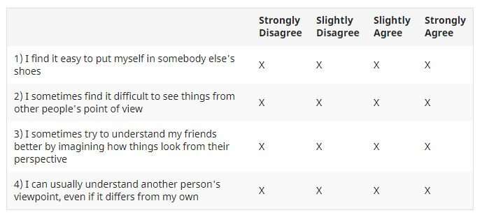
Results from their simple, four-step questionnaire were scored, ranging from 4 to 16 (with 4 indicating poor mind-reading abilities; 16 indicating excellent abilities). The average score for their questionnaire was between 12 and 13. After statistically confirming that the test was measuring the same thing in men and women, they found that females reported better mind-reading than males, whilst also confirming some of the well-reported social challenges faced by the autistic community.
Mind-reading questionnaire developed by researchers at the University of Bath.
Their method, which uses just four questions to assess individuals, is published today, along with their research findings, in the journal Psychological Assessment.
Dr. Punit Shah, senior author of the study and leading expert on social cognitive processing at the University of Bath’s Department of Psychology explained: “We will all undoubtedly have had experiences where we have felt we have not connected with other people we are talking to, where we’ve perceived that they have failed to understand us, or where things we’ve said have been taken the wrong way. Much of how we communicate relies on our understanding of what others are thinking, yet this is a surprisingly complex process that not everyone can do.
“To understand this psychological process, we needed to separate mind-reading from empathy. Mind-reading refers to understanding what other people are thinking, whereas empathy is all about understanding what others are feeling. The difference might seem subtle but is critically important and involves very different brain networks. By focussing carefully on measuring mind-reading, without confusing it with empathy, we are confident that we have just measured mind-reading. And, when doing this, we consistently find that females reported greater mind-reading abilities than their male counterparts.”
Lead researcher, Rachel Clutterbuck, emphasised the clinical importance of the questionnaire. She said: “This new test, which takes under a minute to complete, has important utility in clinical settings. It is not always obvious if someone is experiencing difficulties understanding and responding to others—and many people have learnt techniques which can reduce the appearance of social difficulties, even though these remain.
“This work has great potential to better understand the lived experience of people with mind-reading difficulties, such as those with autism, whilst producing a precise quantitative score that may be used by clinicians to identify individuals who may benefit from interventions.”
Dr. Shah added: “This research has been about understanding more about our mind-reading abilities and providing solutions to those who might struggle, particularly the autistic community. We have created a freely available questionnaire which we hope can help identify people who are experiencing mental difficulties relevant to social situations.”
When I was asked to calculate the total volume of SARS-CoV-2 in the world for the BBC Radio 4 show “More or Less,” I will admit I had no idea what the answer would be. My wife suggested it would be the size of an Olympic swimming pool. “Either that or a teaspoon,” she said. “It’s usually one or the other with these sorts of questions.”
So how to set about calculating an approximation of what the total volume really is? Fortunately, I have some form with these sorts of large-scale back-of-the-envelope estimations, having carried out a number of them for my book The Maths of Life and Death. Before we embark on this particular numerical journey, though, I should be clear that this is an approximation based on the most reasonable assumptions, but I will happily admit there may be places where it can be improved.
So where to start? We’d better first calculate how many SARS-CoV-2 particles there are in the world. To do that, we’ll need to know how many people are infected. (We’ll assume humans rather than animals are the most significant reservoir for the virus.)
According to stats website Our World in Data, half a million people are testing positive for Covid every day. Yet we know that many people will not be included in this count because they are asymptomatic or choose not to get tested, or because widespread testing is not readily available in their country.
The amount of virus that each of the people currently infected will carry around with them (their viral load) depends on how long ago they were infected. On average, viral loads are thought to rise and peak about six days after infection, after which they steadily decline.
Of all the people who are infected now, those who got infected yesterday will contribute a little to the total count. Those who were infected a couple of days ago will contribute a little more. Those infected three days ago a little more still. On average, people infected six days ago will have the highest viral load. This contribution will then decline for people who were infected seven or eight or nine days ago, and so on.
The final thing we need to know is the number of virus particles people harbor at any point during their infection. Since we know roughly how viral load changes over time, it’s enough to have an estimate of the peak viral load. An unpublished study took data on the number of virus particles per gram of a range of different tissues in infected monkeys and scaled up the size of tissue to be representative of humans. Their rough estimates for peak viral loads range from 1 billion to 100 billion virus particles.
Let’s work with the higher end of the estimates so that we get an overestimate of the total volume at the end. When you add up all the contributions to the viral load of each of the three million people who became infected on each of the previous days (assuming this three million rate is roughly constant), then we find that there are roughly two quintillion (2×10¹⁸ or two billion billion) virus particles in the world at any one time.https://www.youtube.com/embed/cQQoLfOXV4E?wmode=transparent&start=0&enablejsapi=1
This sounds like a really big number, and it is. It is roughly the same as the number of grains of sand on the planet. But when calculating the total volume, we’ve got to remember that SARS-CoV-2 particles are extremely small. Estimates of the diameter range from 80 to 120 nanometers. One nanometer is a billionth of a meter. To put it in perspective, the radius of SARS-CoV-2 is roughly 1,000 times thinner than a human hair. Let’s use the average value for the diameter of 100 nanometers in our subsequent calculation.
To work out the volume of a single spherical virus particle we need to use the formula for the volume of a sphere that is, no doubt, on the tip of everyone’s tongue:
V = 4 π r³/3
Assuming a 50 nanometer radius (at the center of the estimated range) of SARS-CoV-2 for the value of r, the volume of a single virus particle works out to be 523,000 nanometres³.
Multiplying this very small volume by the very large number of particles we calculated earlier, and converting into meaningful units gives us a total volume of about 120 milliliters (ml). If we wanted to put all these virus particles together in one place, then we’d need to remember that spheres don’t pack together perfectly.
Close Sphere Packing
If you think about the pyramid of oranges you might see at the grocery store, you’ll remember that a significant portion of the space it takes up is empty. In fact, the best you can do to minimize empty space is a configuration called “close sphere packing” in which empty space takes up about 26 percent of the total volume. This increases the total gathered volume of SARS-CoV-2 particles to about 160ml, easily small enough to fit inside about six shot glasses. Even taking the upper end of the diameter estimate and accounting for the size of the spike proteins all the SARS-CoV-2 still wouldn’t fill a Coke can.
It turns out that the total volume of SARS-CoV-2 was between my wife’s rough estimates of the teaspoon and the swimming pool. It’s astonishing to think that all the trouble, the disruption, the hardship, and the loss of life that has resulted over the last year could constitute just a few mouthfuls of what would undoubtedly be the worst beverage in history.
Summary: Researchers discovered a single gene alteration that may help explain cognitive differences between modern humans and our predecessor, and used that information to develop Neanderthal-like brain organoids in the lab.
As a professor of pediatrics and cellular and molecular medicine at University of California San Diego School of Medicine, Alysson R. Muotri, PhD, has long studied how the brain develops and what goes wrong in neurological disorders. For almost as long, he has also been curious about the evolution of the human brain — what changed that makes us so different from preceding Neanderthals and Denisovans, our closest evolutionary relatives, now extinct?
Evolutionary studies rely heavily on two tools — genetics and fossil analysis — to explore how a species changes over time. But neither approach can reveal much about brain development and function because brains do not fossilize, Muotri said. There is no physical record to study.
So Muotri decided to try stem cells, a tool not often applied in evolutionary reconstructions. Stem cells, the self-renewing precursors of other cell types, can be used to build brain organoids — “mini brains” in a laboratory dish. Muotri and colleagues have pioneered the use of stem cells to compare humans to other primates, such as chimpanzees and bonobos, but until now a comparison with extinct species was not thought possible.
In a study published February 11, 2021 in Science, Muotri’s team catalogued the differences between the genomes of diverse modern human populations and the Neanderthals and Denisovans, who lived during the Pleistocene Epoch, approximately 2.6 million to 11,700 years ago. Mimicking an alteration they found in one gene, the researchers used stem cells to engineer “Neanderthal-ized” brain organoids.
“It’s fascinating to see that a single base-pair alteration in human DNA can change how the brain is wired,” said Muotri, senior author of the study and director of the UC San Diego Stem Cell Program and a member of the Sanford Consortium for Regenerative Medicine. “We don’t know exactly how and when in our evolutionary history that change occurred. But it seems to be significant, and could help explain some of our modern capabilities in social behavior, language, adaptation, creativity and use of technology.”
The team initially found 61 genes that differed between modern humans and our extinct relatives. One of these altered genes — NOVA1 — caught Muotri’s attention because it’s a master gene regulator, influencing many other genes during early brain development. The researchers used CRISPR gene editing to engineer modern human stem cells with the Neanderthal-like mutation in NOVA1. Then they coaxed the stem cells into forming brain cells and ultimately Neanderthal-ized brain organoids.
Brain organoids are little clusters of brain cells formed by stem cells, but they aren’t exactly brains (for one, they lack connections to other organ systems, such as blood vessels). Yet organoids are useful models for studying genetics, disease development and responses to infections and therapeutic drugs. Muotri’s team has even optimized the brain organoid-building process to achieve organized electrical oscillatory waves similar to those produced by the human brain.
The Neanderthal-ized brain organoids looked very different than modern human brain organoids, even to the naked eye. They had a distinctly different shape. Peering deeper, the team found that modern and Neanderthal-ized brain organoids also differ in the way their cells proliferate and how their synapses — the connections between neurons — form. Even the proteins involved in synapses differed. And electrical impulses displayed higher activity at earlier stages, but didn’t synchronize in networks in Neanderthal-ized brain organoids.
According to Muotri, the neural network changes in Neanderthal-ized brain organoids parallel the way newborn non-human primates acquire new abilities more rapidly than human newborns.
“This study focused on only one gene that differed between modern humans and our extinct relatives. Next we want to take a look at the other 60 genes, and what happens when each, or a combination of two or more, are altered,” Muotri said.
“We’re looking forward to this new combination of stem cell biology, neuroscience and paleogenomics. The ability to apply the comparative approach of modern humans to other extinct hominins, such as Neanderthals and Denisovans, using brain organoids carrying ancestral genetic variants is an entirely new field of study.”
To continue this work, Muotri has teamed up with Katerina Semendeferi, professor of anthropology at UC San Diego and study co-author, to co-direct the new UC San Diego Archealization Center, or ArchC.
“We will merge and integrate this amazing stem cell work with anatomic comparisons from several species and neurological conditions to create downstream hypotheses about brain function of our extinct relatives,” Semendeferi said. “This neuro-archealization approach will complement efforts to understand the mind of our ancestors and close relatives, like the Neanderthals.”
Co-authors of the study include: Cleber A. Trujillo, Isaac A. Chaim, Emily C. Wheeler, Assael A. Madrigal, Justin Buchanan, Sebastian Preissl, Allen Wang, Priscilla D. Negraes, and Ryan Szeto, UC San Diego; Edward S. Rice, Nathan K. Schaefer, Ashley Byrne, Maximillian Marin, Christopher Vollmers, Angela N. Brooks, Richard E. Green, UC Santa Cruz; Roberto H. Herai, Pontifícia Universidade Católica do Paraná; Alik Huseynov, Imperial College London; Mariana S.A. Ferraz, Fernando da S. Borges, Alexandre H. Kihara, Universidade Federal do ABC; Jonathan D. Lautz, Stephen E.P. Smith, Seattle Children’s Research Institute and University of Washington; Beth Shapiro, UC Santa Cruz and Howard Hughes Medical Institute; and Gene W. Yeo, UC San Diego, Agency for Science, Technology and Research (Singapore) and National University of Singapore.
Funding for this research came, in part, from the Neanderthal Brain Foundation, National Institutes of Health (grants U19MH1073671, K12GM068524, K01AA026911), Brain and Behavior Research Foundation (NARSAD Independent Investigator Grant), National Science foundation (grant 1754451), Gordon and Betty Moore Foundation (grant GBMF3804), Coordenação de Aperfeiçoamento de Pessoal de Nível Superior (Capes, Brazil), FAPESP (São Paulo Research Foundation, grant 2017/26439-0), CNPq (Brazil’s National Council for Scientific and Technological Development, grants 431000/2016-6, 312047/2017-7) and Loulou Foundation.
Disclosure: Alysson R. Muotri is a co-founder and has equity interest in TISMOO, a company dedicated to genetic analysis and brain organoid modeling focusing on therapeutic applications customized for autism spectrum disorder and other neurological disorders with genetic origins. The terms of this arrangement have been reviewed and approved by the University of California San Diego in accordance with its conflict of interest policies.
Researchers found that Paroxetine not only slows down cartilage degeneration, but also promotes cartilage health in both mice and human cartilage in vitro. Credit: Fadia Kamal, Penn State
A disease of the joints, osteoarthritis affects more than 30 million adults and is the fifth-leading cause of disability in the United States. In a new study, scientists have discovered the cellular pathway that leads to osteoarthritis and have identified a commonly used anti-depressant—paroxetine—that inhibits this pathway. The team found that Paroxetine not only slows down cartilage degeneration, but also promotes cartilage health in both mice and human cartilage in vitro. The drug may be the first-ever treatment for this debilitating, degenerative disease.
“Osteoarthritis destroys joint cartilage and results in pain and disability,” said Fadia Kamal, assistant professor of orthopedics and rehabilitation at Penn State College of Medicine. “Patients live with this pain until their cartilage is extremely degenerated. Unfortunately, an invasive artificial joint replacement surgery is the only treatment orthopedists are currently able to offer. There has been a dire need to identify novel therapeutic targets, approaches or agents that can actively halt or reverse the osteoarthritis disease process.”
In previous research, Kamal and her colleagues found that elevated expression and activity of the enzyme G protein-coupled receptor kinase 2 (GRK2) leads to pathologic cell growth in heart and kidney disease.
Kamal explained that osteoarthritis is similarly driven by pathological growth of cartilage cells, a process called chondrocyte hypertrophy, but how this proliferation occurs had been a mystery. Given their knowledge of the role of GRK2 in heart and kidney disease, Kamal and her team decided to investigate the enzyme in osteoarthritis patients. They found that patients with osteoarthritis or acute injury to the joint had high levels of GRK2 in their cartilage cells, or chondrocytes.
“We discovered a central role for GRK2 in cartilage degeneration, where GRK2 pushes chondrocytes to destroy the cartilage matrix surrounding them instead of replenishing and maintaining it.” said Kamal. “In other words, the cells receive a bad signal to destroy cartilage.”
The researchers confirmed the role of GRK2 in cartilage degeneration in two experiments: in one, they performed a genetic deletion of GRK2 from cartilage cells in mice, and in the other, they administered paroxetine—an FDA-approved selective serotonin reuptake inhibitor (SSRI) that is a potent GRK2 inhibitor—to the mice. In both cases, they found that not only did GRK2 deletion prevent chondrocyte hypertrophy and halt osteoarthritis progression, but it also promoted cartilage regeneration.
“We found that paroxetine could return cartilage cells back to a normal state and preserve the cartilage surface,” said Kamal.
In other experiments with cultured human osteoarthritic cartilage, obtained from patients undergoing knee replacement surgery, the team also confirmed the ability of paroxetine to mitigate chondrocyte hypertrophy and cartilage degradation.
The results will appear on Feb. 10 in the journal Science Translational Medicine.
“Our findings present elevated GRK2 signaling in chondrocytes as a driver of chondrocyte hypertrophy and cartilage degradation and identify paroxetine as a disease-modifying drug for OA treatment,” said Kamal. “This is important given that around 80% of the U.S. population will develop radiographic evidence of osteoarthritis by age 65 and with the growing prevalence of osteoarthritis risk factors, such as obesity and diabetes, osteoarthritis will likely carry an even greater burden in the future.”
The team is currently seeking approval from the FDA for a new trial of this drug to treat osteoarthritis.
“If this trial works, we will have found a new solution to an age-old problem of joints in the body wearing out because of cartilage destruction and loss,” said Kamal. “We hope to intervene with this disease-modifying treatment for the benefit of our patients.”
Adults with type 2 diabetes and overweight or obesity assigned a once-monthly monoclonal antibody infusion experienced a marked decrease in fat mass and gains in muscle vs. those assigned placebo, according to findings from a phase 2 study.
Bimagrumab, a human monoclonal antibody that blocks activin type II receptors and stimulates muscle growth, was not initially investigated as an obesity treatment, Steven B. Heymsfield, MD, FTOS, professor in the department of metabolism and body composition at Pennington Biomedical Research Center, Louisiana State University, told Healio. The drug was initially heralded as a potential breakthrough therapy for people with sporadic inclusion body myositis, a rare muscle-wasting disease. However, the drug did not meet its primary endpoint in a phase 2b/3 trial, Novartis announced in a press release in 2016.
“When researchers did the preclinical work, there was absolutely no signal on adipose tissue; it was all muscle growth,” Heymsfield said in an interview. “They had no reason to suspect it, because [activin type II receptors] are mainly muscle. When they did first-in-man studies, they did see some adipose tissue signal and conducted a proof-of-concept study to see if adipose tissue effects were significant. That is what prompted the current investigation — a phase 2 trial with body fat as the primary endpoint. As of today, there is not a clear mechanism.”
Study design
Heymsfield and colleagues analyzed data from 75 adults with type 2 diabetes with overweight or obesity, defined as a BMI between 28 kg/m² and 40 kg/m² (mean age, 60 years; mean BMI 32.9 kg/m²; mean body weight, 93.6 kg; mean fat mass, 35.4 kg; mean HbA1c, 7.8%). The trial was conducted from February 2017 to May 2019. Researchers randomly assigned participants an IV infusion of bimagrumab (10 mg/kg up to 1,200 mg in 5% dextrose solution; n = 37; 62.2% women) or placebo (5% dextrose solution; n = 38; 77.3% women) every 4 weeks for 48 weeks. Both groups received diet and exercise counseling. The primary endpoint was least square mean change from baseline to week 48 in total body fat mass as measured by DXA; secondary and exploratory endpoints were lean mass, waist circumference, HbA1c and body weight changes from baseline to week 48.
Fat mass vs. body weight
At week 48, participants in the bimagrumab groups experienced a mean –20.5% loss in fat mass (mean, –7.5 kg; 80% CI, –8.3 to –6.6) vs. a mean –0.5% reduction for those in the placebo group (–0.18 kg; 80% CI, –0.99 to –0.63).
Participants assigned bimagrumab also experienced a mean gain of 3.6% in lean mass (mean, 1.7 kg; 80% CI, 1.1-2.3) compared with a mean –0.8% reduction in lean mass for the placebo group (mean, –0.4 kg; 80% CI, –1 to 0.1).
Waist circumference decreased by a mean of 9 cm in the bimagrumab group vs. a 0.5 cm gain in the placebo group (P < .001), and HbA1c fell by 0.76 percentage points in the bimagrumab group vs. 0.04 percentage points in the placebo group (P = .005).
Weight loss was also greater in the bimagrumab group vs. placebo (mean, –5.9 kg vs. –0.8 kg; P < .001).
Bimagrumab’s safety and tolerability profile was consistent with prior studies. Mild diarrhea and muscle spasms were the most commonly reported adverse events in the bimagrumab group; one patient in the bimagrumab group developed pancreatitis.
“What surprised me the most was the magnitude of the effects on body fat,” Heymsfield said. “The effect is real; this is not a one-off. People lost 7.5 kg of fat, or almost 16 to 20 pounds of fat. That is significant fat loss, particularly for people with diabetes, who tend not to respond very well to anti-obesity treatment.”
There is excitement about bimagrumab and the possible mechanism behind the new findings; however, next steps in the drug’s pipeline are unclear, Heymsfield said. Novartis opted to license the drug and has not disclosed who the licensee is, he added.
“It’s not dead,” Heymsfield said of the therapy. “To be candid, the diabetes space is pretty crowded. You can take metformin for a penny a day. Monoclonal antibodies are also expensive.”
Researchers also noted a signal for elevated pancreatic enzymes, the origin or significance of which is unclear, he said.
“The real future of this drug involves figuring out the mechanism, working through that and finding targets that are druggable,” Heymsfield said. “This study demonstrates the beguiling nature of weight changes. These people lost more fat than body weight. You cannot always rely on weight as an index of efficacy.”
People go to bed later and sleep fewer hours the night before a full moon, and menstrual cycles seem to temporarily synchronize with moon cycles, scientists have found in two new studies.
Does the full moon change how we sleep? Does it synchronize with menstrual cycles?
What might sound like old-school myths might actually hold some truth. People go to bed later and sleep fewer hours before a full moon and menstrual cycles seem to temporarily synchronize with moon cycles, scientists have found in two new studies.
Throughout history, humans have connected our daily lives to the changing skies, specifically the changing faces of the moon. Lore surrounding the moon’s phases has ranged from full moons inciting werewolves to the moon’s cycle affecting how we feel and our day-to-day moods.
But, strangely, a couple of these tall tales seem to have roots in real science.
In a study published January 27 in the journal Science Advances, a team of scientists from the University of Washington, the National University of Quilmes in Argentina and Yale University show how sleep cycles seem to change with the lunar cycle.
They found that, in the days leading up to a full moon, people tend to go to sleep later and sleep for fewer hours. For this work, the team studied college students in the city of Seattle, Washington, and also with those living in indigenous communities in northern Argentina, two different environments where there is a variety in individual access to electricity because of how artificial light might affect the participants.
Using sleep-monitoring wrist devices, they studied 98 individuals living in three Toba-Qom indigenous communities in Formosa, Argentina and additionally used sleep data from 464 college students in the Seattle area (the data from the college students was originally collected for a separate study).
The team found that, while the connection between sleep cycles and lunar cycles is a bit more obvious in communities without electricity access, the connection still seems to be present in areas with electricity as well.
“We see a clear lunar modulation of sleep, with sleep decreasing and a later onset of sleep in the days preceding a full moon,” lead author Horacio de la Iglesia, a professor of biology at the University of Washington, said in a statement. “And although the effect is more robust in communities without access to electricity, the effect is present in communities with electricity, including undergraduates at the University of Washington.”
In these groups, they showed that the nights leading up to a full moon was when people slept the least and went to bed the latest. These nights also had more light in the night sky after dusk as the waxing moon got brighter.
“We hypothesize that the patterns we observed are an innate adaptation that allowed our ancestors to take advantage of this natural source of evening light that occurred at a specific time during the lunar cycle,” study author Leandro Casiraghi, a University of Washington postdoctoral researcher in the biology department.
Menstrual and lunar cycles
Sleep cycles aren’t the only human function that seems to be affected by the moon, scientists are finding. This is not a new notion. In fact, for a long time, people have suggested that there is a connection between lunar and menstrual cycles, some myths even suggesting that fertility and lunar cycles have some sort of connection, a controversial tale.
In a separate study, also published today in Science Advances, researchers showed that, while all of the myths surrounding this connection might not hold up, there could be some link between menstrual cycles and moon cycles.
By analyzing menstrual cycle records that 22 women kept for up to 32 years. They examined long-term data on menstrual cycle onset with data averaging a length of 15 years and including information from women both under and over age 35. They compared this data with fluctuations in the lunar cycles to see how the two lined up.
They found that, of the women who participated, those whose menstrual cycles last longer than 27 days showed “intermittently synchronized with cycles that affect the intensity of moonlight,” according to a statement. The team determined that this synchronization was slowly lost over time as the participants grew older, and found that the link was lessened with increased exposure to artificial light.
More specifically, they concluded that “menstrual cycles also aligned with the tropical month (the 27.32 days it takes the moon to pass twice through the same equinox point) 13.1% of the time in women 35 years and younger and 17.7% of the time in women over 35, suggesting that menstruation is also affected by shifts in the moon’s gravimetric forces,” according to the statement.
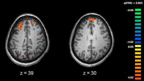
Functional magnetic resonance imaging (fMRI) and other brain imaging technologies allow for the study of differences in brain activity in people diagnosed with schizophrenia. The image shows two levels of the brain, with areas that were more active in healthy controls than in schizophrenia patients shown in orange, during an fMRI study of working memory. Credit: Kim J, Matthews NL, Park S./PLoS One.
People with schizophrenia, a mental disorder that affects mood and perception of reality, are almost three times more likely to die from the coronavirus than those without the psychiatric illness, a new study shows. Their higher risk, the investigators say, cannot be explained by other factors that often accompany serious mental health disorders, such as higher rates of heart disease, diabetes, and smoking.
Led by researchers at NYU Grossman School of Medicine, the investigation showed that schizophrenia is by far the biggest risk factor (2.7 times increased odds of dying) after age (being 75 or older increased the odds of dying 35.7 times). Male sex, heart disease, and race ranked next after schizophrenia in order.
“Our findings illustrate that people with schizophrenia are extremely vulnerable to the effects of COVID-19,” says study lead author Katlyn Nemani, MD. “With this newfound understanding, health care providers can better prioritize vaccine distribution, testing, and medical care for this group,” adds Nemani, a research assistant professor in the Department of Psychiatry at NYU Langone Health.
The study also showed that people with other mental health problems such as mood or anxiety disorders were not at increased risk of death from coronavirus infection.
Since the beginning of the pandemic, experts have searched for risk factors that make people more likely to succumb to the disease to bolster protective measures and allocate limited resources to people with the greatest need. Although previous studies have linked psychiatric disorders in general to an increased risk of dying from the virus, the relationship between the coronavirus and schizophrenia specifically has remained unclear. A higher risk of mortality was expected among those with schizophrenia, but not at the magnitude the study found, the researchers say.
The new investigation is publishing Jan. 27 in the journal JAMA Psychiatry. Researchers believed that other issues such as heart disease, depression, and barriers in getting care were behind the low life expectancy seen in schizophrenia patients, who on average die 15 years earlier than those without the disorder. The results of the new study, however, suggest that there may be something about the biology of schizophrenia itself that is making those who have it more vulnerable to COVID-19 and other viral infections. One likely explanation is an immune system disturbance, possibly tied to the genetics of the disorder, says Nemani.
For the investigation, the research team analyzed 7,348 patient records of men and women treated for COVID-19 at the height of the pandemic in NYU Langone hospitals in New York City and Long Island between March 3 and May 31, 2020. Of these cases, they identified 14 percent who were diagnosed with schizophrenia, mood disorders, or anxiety. Then, the researchers calculated patient death rates within 45 days of testing positive for the virus.
They note that this large sample of patients who all were infected with the same virus provided a unique opportunity to study the underlying effects of schizophrenia on the body.
“Now that we have a better understanding of the disease, we can more deeply examine what, if any, immune system problems might contribute to the high death rates seen in these patients with schizophrenia,” says study senior author Donald Goff, MD. Goff is the Marvin Stern Professor of Psychiatry at NYU Langone.
Goff, also the director of the Nathan S. Kline Institute for Psychiatric Research at NYU Langone, says the study investigators plan to explore whether medications used to treat schizophrenia, such as antipsychotic drugs, may play a role as well.
He cautions that the study authors could only determine the risk for patients with schizophrenia who had access to testing and medical care. Further research is needed, he says, to clarify how dangerous the virus may be for those who lack these resources. Goff is also the vice chair for research in the Department of Psychiatry at NYU Langone.
Hormones during pregnancy. Depressed expectant lady closing her face and crying, grey studio background
New research from the University of Iowa and University Hospitals Cleveland Medical Center demonstrates that offspring can be protected from the effects of prenatal stress by administering a neuroprotective compound during pregnancy.
Working in a mouse model, Rachel Schroeder, a student in the UI Interdisciplinary Graduate Program in Neuroscience, drew a connection between the work of her two mentors, Hanna Stevens, MD, PhD, UI associate professor of psychiatry and Ida P. Haller Chair of Child and Adolescent Psychiatry, and Andrew A. Pieper, MD, PhD, a former UI faculty member, now Morley-Mather Chair of Neuropsychiatry at Case Western Reserve University and Investigator and Director of the Neurotherapeutics Center at The Harrington Discovery Institute at University Hospitals.
Stevens’s lab studies the long-lasting impact of stress during pregnancy, which can lead to neuropsychiatric impairment in offspring during early life and in adulthood. Pieper’s lab focuses on discovery of neuroprotective treatments, exemplified by the pharmacologic agent used here, known as P7C3-A20, which has previously been shown to protect the adult brain from injury.
Schroeder spent time in both labs early in her graduate school career and was inspired to bring the two lines of research together in her own work, investigating the potential impact that P7C3-A20 might have in protecting the embryonic brain during adverse events in pregnancy. Her work is the first to explore the therapeutic potential of prenatal exposure to P7C3 compounds.
“Prenatal stress increases the risk for offspring to have neurodevelopmental problems,” Schroeder said. “We wanted to know whether the P7C3-A20 compound protected the embryonic brain from damage. Our results show that offspring are protected from the negative effects of stress when the mothers are treated with P7C3-A20 during the same time.”
Previous work by Pieper’s lab has shown that P7C3-A20 enables nerve cells to maintain normal levels of an energy molecule known as nicotinamide adenine dinucleotide (NAD+), under toxic or injury conditions that are otherwise overwhelming and energy-depleting for the cell.
Schroeder’s research showed that chronic prenatal stress in mice disrupted the embryonic brain’s NAD+-synthesis machinery, which led to degeneration of nerve cell axons, learning deficits, and depression-like behavior when the offspring reached adulthood. Schroeder demonstrated that when prenatally-stressed pregnant mice were simultaneously treated with P7C3-A20, their offspring were protected from these negative effects.
“By stabilizing critical NAD+-producing mechanisms, we enabled the developing embryonic brain to continue developing normally despite the stress,” Schroeder said.
“Though there are many challenges associated with administering medicines during pregnancy, Rachel Schroeder’s discovery represents an exciting move forward in understanding how prenatal stress harms the brain, and strategies for protecting the developing embryo.” said Pieper, who is also Psychiatrist at the Louis Stokes VA Medical Center in Cleveland.
This study represents an important proof of concept for a new approach to early prevention of neuropsychiatric problems, Stevens said. “Neuropsychiatric problems are the most common chronic illnesses of young people, which means we need many more ways to protect the brain as it develops. Our lab is focused on mechanisms of brain development prenatally, a critical time when we could make a difference.”
In addition to Schroeder, Pieper and Stevens, the research team included Lynn Nguyen and Alexandra Loren in the Stevens Lab at UI; Preethy Sridharan, Coral J. Cintrón-Pérez, and Edwin Vázquez-Rosa in the Pieper Lab at the Harrington Discovery Institute; and Noelle S. Williams and Kavitha P. Kettimuthu at the University of Texas Southwestern Medical Center.
###
Funding was provided by the Brockman Foundation, the Elizabeth Ring Mather & William Gwinn Mather Fund, S. Livingston Samuel Mather Trust, G.R. Lincoln Family Foundation, Wick Foundation, Gordon & Evie Safran, the Leonard Krieger Fund of the Cleveland Foundation, the Maxine and Lester Stoller Parkinson’s Research Fund, the Louis Stokes VA Medical Center resources and facilities, Project 19PABH134580006-AHA/Allen Initiative in Brain Health and Cognitive Impairment, a Junior Research Program of Excellence from the Roy J. Carver Charitable Trust, Nellie Ball Trust, NIH grant R01 MH122485-01, UI Environmental Health Science Research Center and the UI Graduate College.
About University Hospitals / Cleveland, Ohio Founded in 1866, University Hospitals serves the needs of patients through an integrated network of 19 hospitals, more than 50 health centers and outpatient facilities, and 200 physician offices in 16 counties throughout northern Ohio. The system’s flagship academic medical center, University Hospitals Cleveland Medical Center, located in Cleveland’s University Circle, is affiliated with Case Western Reserve University School of Medicine. The main campus also includes University Hospitals Rainbow Babies & Children’s Hospital, ranked among the top children’s hospitals in the nation; University Hospitals MacDonald Women’s Hospital, Ohio’s only hospital for women; University Hospitals Harrington Heart & Vascular Institute, a high-volume national referral center for complex cardiovascular procedures; and University Hospitals Seidman Cancer Center, part of the NCI-designated Case Comprehensive Cancer Center. UH is home to some of the most prestigious clinical and research programs in the nation, including cancer, pediatrics, women’s health, orthopedics, radiology, neuroscience, cardiology and cardiovascular surgery, digestive health, transplantation and urology. UH Cleveland Medical Center is perennially among the highest performers in national ranking surveys, including “America’s Best Hospitals” from U.S. News & World Report. UH is also home to Harrington Discovery Institute at University Hospitals – part of The Harrington Project for Discovery & Development. UH is one of the largest employers in Northeast Ohio with 28,000 physicians and employees. Advancing the Science of Health and the Art of Compassion is UH’s vision for benefitting its patients into the future, and the organization’s unwavering mission is To Heal. To Teach. To Discover. Follow UH on LinkedIn, Facebook @UniversityHospitals and Twitter @UHhospitals. For more information, visit UHhospitals.org.
About Harrington Discovery Institute The Harrington Discovery Institute at University Hospitals in Cleveland, OH—part of The Harrington Project for Discovery & Development—aims to advance medicine and society by enabling our nation’s most inventive scientists to turn their discoveries into medicines that improve human health. The institute was created in 2012 with a $50 million founding gift from the Harrington family and instantiates the commitment they share with University Hospitals to a Vision for a ‘Better World’. For more information, visit: HarringtonDiscovery.org.
Case Western Reserve University is one of the country’s leading private research institutions. Located in Cleveland, we offer a unique combination of forward-thinking educational opportunities in an inspiring cultural setting. Our leading-edge faculty engage in teaching and research in a collaborative, hands-on environment. Our nationally recognized programs include arts and sciences, dental medicine, engineering, law, management, medicine, nursing and social work. About 5,100 undergraduate and 6,700 graduate students comprise our student body. Visit case.edu to see how Case Western Reserve thinks beyond the possible.
Louis Stokes Cleveland VA Medical Center is the hub of VA Northeast Ohio Healthcare System, providing and coordinating primary, acute and specialty care. Focusing on treating the whole Veteran through health promotion and disease prevention, VA Northeast Ohio Healthcare System delivers comprehensive, seamless health care and social services for more than 112,000 Veterans at 18 locations across Northeast Ohio. VA Northeast Ohio Healthcare System contributes to the future of medicine through education, training and research programs. For more information visit www.cleveland.va.gov.