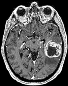
A new pathway that is used by cancer cells to infiltrate the brain has been discovered by a team of Canadian and American research groups led by the Singh Lab at McMaster University. The research also reveals a new therapy that shows promise in blocking and killing these tumors.
The research, published in Nature Medicine on Aug. 2, 2024, offers new hope and potential treatments for glioblastoma, the most aggressive form of brain cancer.
With existing treatments like surgery, radiation therapy and chemotherapy, the tumors often return, and patient survival is limited to only a few months. With this new treatment, the returning cancer cells were destroyed at least 50% of the time in two of the three diseases tested in preclinical animal models.
To discover the pathway cancer cells use to infiltrate the brain, researchers used large-scale gene editing technology to compare gene dependencies in glioblastoma when it was initially diagnosed and after it returned following standard treatments. By doing this, researchers discovered a new pathway used for axonal guidance—a signaling axis that helps establish normal brain architecture—that can become overrun by cancer cells.
“In glioblastoma, we believe that the tumor hijacks this signaling pathway and uses it to invade and infiltrate the brain,” says co-senior author Sheila Singh, professor with McMaster’s Department of Surgery and director of the Center for Discovery in Cancer Research. The research was also co-led by Jason Moffat, head of the Genetics and Genome Biology program at The Hospital for Sick Children (SickKids).
“If we can block this pathway, the hope is that we can block the invasive spread of glioblastoma and kill tumor cells that cannot be removed surgically,” says Singh.
Promising new therapeutic
To stop the invasion of cancer cells, researchers targeted the hijacked signaling pathway using different strategies including a drug developed by John Lazo’s group at the University of Virginia, and also by developing a new therapy with help from Kevin Henry and Martin Rossotti at the National Research Council Canada using CAR T cells to target the pathway in the brain.
They honed in on a protein called Roundabout Guidance Receptor 1 (ROBO1) that helps guide certain cells, similar to a GPS.
“We created a type of cell therapy where cells are taken from a patient, edited and then put back in with a new function. In this case, the CAR T cells were genetically edited to have the knowledge and ability to go and find ROBO1 on tumor cells in animal models,” says lead author Chirayu Chokshi, a former Ph.D. student who worked alongside Singh at McMaster University.
Singh and Chokshi say the treatment can also apply to other invasive brain cancers. In the study, researchers examined models for three different types of cancer including adult glioblastoma, adult lung-to-brain metastasis, and pediatric medulloblastoma. In all three models, treatment led to a doubling of survival time. In two of the three diseases, it led to tumor eradication in at least 50% of the mice.
“In this study, we present a new CAR T therapy that is showing very promising preclinical results in multiple malignant brain cancer models, including recurrent glioblastoma. We believe our new CAR T therapy is poised for further development and clinical trials,” Singh says.
Work on the study was performed with samples derived from patients treated by neurosurgeons with Hamilton Health Sciences. Proteomics discovery which helped to elucidate the new glioblastoma targets was done in collaboration with Thomas Kilinger at Princess Margaret Cancer Center and University of Toronto.
The research was made possible through collaboration with the National Research Council Canada, University of Virginia, University of Pittsburgh and the Princess Margaret Cancer Center.
Young american football player running back breaking away from an attempted tackle. All logos and trademarks from uniforms, helmets and cleats have been removed in Photoshop
Summary: Researchers discover playing football for more than 11 years is tied to less white matter in the brain, could lead to poor impulse control and thinking problems.
The degenerative brain disease known as CTE, or chronic traumatic encephalopathy, has become a specter haunting football. One-time stars—like the late NFL defensive backs Irv Cross and Dave Duerson and the Hall of Fame center Mike Webster—who were all once heralded for their swaggering on-field heroics, later found themselves condemned to far less glamorous retirements, stuck with years of progressively declining brain health, plagued by forgetfulness, disordered thinking, and poorly regulated emotions.
Now, a new study led by the Boston University CTE Center suggests the shots players take on the path to fame and glory may have a wider impact on their brains than previously known. Researchers found repetitive blows to the head may also lead to less white matter in the brain, potentially causing impulsive behavior and other thinking-related problems, whether or not someone has CTE. The research, published in Brain Communications, showed those who start playing tackle football at an early age or play it for more than 11 years are at greater risk.
“Just because you aren’t diagnosed with CTE doesn’t mean there isn’t something structurally damaged in the brain,” says neuropathologist Thor D. Stein, a BU Chobanian & Avedisian School of Medicine associate professor of pathology and laboratory medicine. “Damage to the white matter may help explain why football players appear more likely to develop cognitive and behavioral problems later in life, even in the absence of CTE.”
Damage from Football’s Repeated Hits
White matter is the brain’s cabling, made up of axons, or nerve fibers, that connect its billions of cells. It accounts for about half of the human brain’s volume—without it, our cells (the gray matter) wouldn’t be able to communicate with each other.
“A lot of neuroscience and degenerative disease study is focused on the neurons or cells themselves, but increasingly people are recognizing that there can be damage to the connections,” says Stein, leader of the BU Alzheimer’s Disease Research Center’s neuropathology core and a staff neurologist at two Boston-area Department of Veterans Affairs’ healthcare systems. “The cell itself might look okay, but its connection is not intact—and that was what we wanted to look at in this study.”
To dig into the effect of repeated hits to the head on these connections, the researchers analyzed the brains of 205 amateur and professional football players. All had asked that their brains be donated to the BU-hosted UNITE Brain Bank, which holds more than 1,200 brains, after their deaths. A majority of the former players—75.9 percent—had reportedly been functionally impaired and, the researchers found, many (but not all) also had CTE.
For the study, Stein and his colleagues split themselves into two groups, blinded—or working independently—from each other. One group conducted a pathological examination of the brains, peering at samples through microscopes and dissecting white matter tissue to test protein levels. The second group evaluated medical records and interviewed family members about symptoms.
Stein was part of the pathological team. He concentrated his efforts on investigating myelin, a membrane of lipids and proteins that wraps around and strengthens the brain’s cabling—like the plastic casing around insulated wire. Using biochemical tests called immunoassays, he measured the levels of two myelin proteins, myelin-associated glycoprotein (MAG) and proteolipid protein 1 (PLP). “How much of these proteins are present is a proxy of the integrity of the white matter,” says Stein. Less myelin, less efficient connections between brain cells.
The researchers targeted the frontal lobe, the part of the brain that controls many executive functions, from memory and attention to planning and self-control. It’s also on the front lines when it comes to football hits and concussion impacts. They found that the more years someone played football, the less PLP they had; those who played for more than 11 years had less PLP and MAG than those with shorter careers. They also discovered that donors who started playing tackle football earlier had lower PLP levels. Stein suspects that young, developing brains are especially susceptible to damage from football’s repeated hits.
“Maybe young folks playing at an early age, their connections might be particularly susceptible to damage,” he says. “We found if you started at a younger age, you were more likely to have less of these white matter–associated proteins decades later in life.”
During their lifetimes, the former players probably struggled to plan their days, control their emotions, and understand the consequences of their actions, says Stein. “In our study, we found that, in those over 50 years of age, lower measures of white matter were associated with an impaired ability to perform normal activities of daily living, such as paying bills, shopping, and cooking, as well as with more impulsive behavior.”
Assess the Risk of Contact Sports
The latest study should allow researchers to give families some closure—by explaining what caused their loved ones’ sliding brain health. The research could also provide a foundation for helping future patients.
“These results suggest that existing tests that measure white matter injury during life, including imaging and blood tests, may help to clarify potential causes of changes in behavior and cognition in former contact sport athletes,” says Michael L. Alosco, a lead author on the study and a Chobanian & Avedisian School of Medicine associate professor of neurology. “We can also use these tests to better understand how repeated hits to the head from football and other sports lead to long-term injury to the white matter.”
Stein hopes their work will also help people better assess the risks of playing football, along with other contact sports.
“There’s a cumulative risk—the more you play, the more your risk is increased,” says Stein, who backs the Concussion Legacy Foundation’s Flag Football Under 14 campaign. “One message we try to get across is you don’t need to be playing tackle football at a very young age—if you can just shrink those cumulative years of play down a little bit, you can make a really big impact on brain health. This study is more evidence of that.”
The Lancet Commission identified high cholesterol and vision loss as new risk factors for dementia.
The commission outlined 13 recommendations for individuals and governments to prevent dementia.
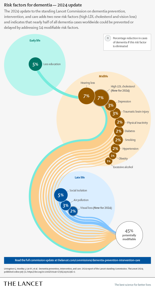
PHILADELPHIA — Tackling 14 risk factors for dementia beginning in childhood could prevent or delay nearly half of cases worldwide, according to a report from the Lancet Commission presented at the Alzheimer’s Association International Conference.
These include two risk factors — high cholesterol and vision loss — newly identified by the commission on dementia prevention, intervention and care.
An estimated 57 million people were living with dementia in 2019, Gill Livingston, MD, a professor of psychiatry at University College London, and colleagues wrote in the report. This number is expected to increase to 153 million by 2050, highlighting the need for risk reduction strategies.
The new report is an update to the commission’s 2020 report. Members of the commission adopted a triangulation framework that prioritized systematic reviews and meta-analyses. They also conducted new meta-analyses when necessary.
The researchers said their review supports the 12 potentially modifiable risk factors that were identified in the 2020 report: air pollution (RR = 1.1; 95% CI, 1.1-1.1); depression (RR = 2.2; 95% CI, 1.7-3); diabetes (RR = 1.7; 95% CI, 1.6-1.8); excessive alcohol use (RR = 1.2; 95% CI, 1-1.5); hearing loss (RR = 1.4; 95% CI, 1-1.9); hypertension (RR = 1.2; 95% CI, 1.1-1.4); lower education level (RR = 1.6; 95% CI, 1.3-2); obesity (RR = 1.3; 95% CI, 1-1.7); physical inactivity (RR = 1.2; 95% CI, 1.2-1.3); smoking (RR = 1.3; 95% CI, 1.2-1.4); social isolation (RR = 1.6; 95% CI, 1.3-1.8); and traumatic brain injury (RR = 1.7; 95% CI, 1.4-1.9).
The evidence also supports the addition of high LDL cholesterol (RR = 1.3; 95% CI, 1.3-1.4) and vision loss (RR = 1.5; 95% CI, 1.4-1.6).
If these 14 risk factors are eliminated, “nearly half of dementias could theoretically be prevented,” Livingston and colleagues wrote.
This has important implications for physicians, particularly family physicians, Livingston told Healio. She noted that diabetes, excessive alcohol use, hearing impairment, high LDL, hypertension, obesity, vision loss and smoking account for about one-quarter of all dementias.
“If we add depression, traumatic brain injury and physical inactivity, which family physicians also advise on, then it is a third of dementias,” she said. “Their active vigilance and advice potentially make a huge difference.”
Based on their findings, the researchers outlined 13 recommendations for individuals and governments to prevent dementia:
ensure children have access to good-quality education and encourage individuals in midlife to participate in “cognitively stimulating activities;”
reduce harmful noise exposure and make hearing aids accessible to those with hearing impairment;
treat depression;
promote helmets and other head protection during contact sports and when riding bicycles;
encourage exercise;
reduce smoking through education and by implementing policies that aim to control the cost of cigarettes;
prevent or reduce high blood pressure;
diagnose and treat high LDL;
maintain a healthy weight and treat obesity early;
reduce excessive alcohol use through price control and raising awareness about the risks of overconsumption;
reduce social isolation by encouraging activities and living with other people, prioritizing an “age-friendly and supportive community, environments and housing”;
ensure access to vision loss screening and treatment; and
decrease air pollution exposure.
“Although addressing risk factors at an early stage of life is desirable, there is also benefit from tackling risk throughout life; it is never too early or too late to reduce dementia risk,” Livingston and colleagues wrote.
References:
Livingston G, et al. Lancet standing commission on dementia prevention, intervention and care. Scientific advances in the 2024 commission. Presented at: Alzheimer’s Association International Conference; July 28-Aug. 1, 2024; Philadelphia.
The Lancet: Nearly half of dementia cases could be prevented or delayed by tackling 14 risk factors starting in childhood, including two new risks — high cholesterol and vision loss. www.eurekalert.org/news-releases/1052982. Published July 31, 2024. Accessed July 31, 2024.
Dementia risk reduction is an important area of research. In this latest Lancet Commission report, they’re adding two more risk factors — high cholesterol and vision loss — to the list and calculating that, together, these 14 factors could account for around half of all worldwide cases of dementia. This illustrates the importance of our awareness of these types of risk factors.
It’s important for our understanding that these reports are not just informed by epidemiological studies, but further interventional studies. One of those, which is ongoing, is the U.S. POINTER study. Recruiting is complete, but evaluating the results of the study is still underway. They will be reported next year at AAIC 2025 in Toronto. This study is looking at multiple risk factors, including modifying diet, exercise, cognitive activities, social engagement and the management of heart health status, and whether these factors in combination can protect cognitive health.
Of note, for right now, the Alzheimer’s Association provides 10 Healthy Habits for Your Brain, which is a great resource for anybody who is thinking about their risk.
Claire Sexton, DPhil
Senior director of scientific programs and outreach
Alzheimer’s Association
Disclosures: Sexton reports no relevant financial disclosures.
MONDAY, July 22, 2024 (HealthDay News) — Dogs can sniff out whether a human is stressed or relaxed, new research suggests, and that sensory feedback appears to influence canine emotions and choices.
The dog doesn’t even have to know the human well to interpret odor in this way, the British researchers noted.
“Dog owners know how attuned their pets are to their emotions, but here we show that even the odor of a stressed, unfamiliar human affects a dog’s emotional state, perception of rewards and ability to learn,” said study author Dr. Nicola Rooney. She’s a senior lecturer in wildlife and conservation at Bristol Veterinary School in Bristol, England.
“Working dog handlers often describe stress traveling down the lead, but we’ve also shown it can also travel through the air,” she said in a university news release.
Her team published its findings July 22 in the journal Scientific Reports.
As the Bristol team noted, research has long pointed to scent as an important but perhaps under-appreciated form of emotional communication between people.
Rooney’s group wondered if dogs, with olfactory senses that are so much more sophisticated than humans, might catch human emotions through smell, as well, and act accordingly.
They constructed an elaborate experiment to find out. First, they trained dogs in a simple task: If a bowl was placed in one location, it invariably contained food. But if it was placed in a separate location, no food was present.
For obvious reasons, the dogs soon became more eager to trot over to bowls in the “have” spot than the “have not” location.
But what if the bowl was placed between these locations?
If the pooch ambled quickly over to this ambiguous, mid-range bowl, the researchers considered that the dog was in an “optimistic” frame of mind (“maybe there’s food in that bowl!”).
If the dog was more hesitant about heading towards the bowl, that reflected a more “pessimistic” attitude (“The bowl’s in the wrong spot, probably no food there”).
Next, the 18 dogs recruited for the experiment were exposed to sweat and breath samples from humans who’d been in either a stressed or relaxed state of mind (a math test versus listening to soothing music).
When dogs smelled the “stressed” human odors, they were visibly less eager to head towards the ambiguously placed bowl, suggesting an emotional downturn towards pessimism, the researchers said.
“This ‘pessimistic’ response reflects a negative emotional state and could possibly be a way for the dog to conserve energy and avoid disappointment,” the researchers reasoned.
However, this “downer” effect was not seen when the dogs were exposed to a “relaxed” odor sample from a human.
According to Rooney, the new findings have real-world applications.
“Understanding how human stress affects dogs’ well-being is an important consideration for dogs in kennels and when training companion dogs and dogs for working roles such as assistance dogs,” she said.
The psychedelic drug causes some lasting changes to the communication pathways that connect distinct brain regions. This heat map shows how patterns of resting brain activity (blue and green) change when psilocybin is taken (red and yellow), then return to normal as the drug wears off. Credit: Sara Moser/Washington University
Taking psilocybin, the hallucinogenic compound found in magic mushrooms, temporarily resets entire networks of neurons in the brain that are responsible for controlling a person’s sense of time and self, finds a study that repeatedly imaged the brains of seven volunteers before, during and after they took a massive dose of the drug.
The findings, published in Nature on 17 July1, could offer insights into why the compound might have a therapeutic effect on some neurological conditions.
Researchers “saw such massive changes induced by psilocybin” that some study participants’ brain-network patterns resembled those of a different person entirely, says Shan Siddiqi, a psychiatric neuroscientist at Harvard School of Medicine in Boston, Massachusetts. “I’ve never seen an effect this strong.”
Most of these changes lasted for a few hours, but one key link between different parts of the brain remained disrupted for weeks.
Psychedelic medicine
Psilocybin is one of several psychedelic drugs, including LSD, ketamine and MDMA (also known as ecstasy), that are being investigated as therapies for conditions such as depression and post-traumatic stress disorder. Despite promising data that have sped treatments towards approval, researchers still don’t fully understand the mechanism that underlies their therapeutic effects.
Many studies have investigated how psychedelics affect individual cells, but Joshua Siegel, a systems neuroscientist at the Washington University School of Medicine in St. Louis, Missouri, took a broader approach to look at how psilocybin affects networks of neurons across the whole brain.
Siegel and his colleagues tracked activity in the brains of seven healthy adults before, during and after they took a high dose of psilocybin. The researchers used functional magnetic resonance imaging (fMRI) to obtain images of blood-flow changes in different parts of the brain — a proxy used to measure how groups of neurons across the brain communicate with one another.
The researchers compared these fMRI scans with images of the same participants’ brains when they did not take any drug or when they took a stimulant. They found that psilocybin caused groups of neurons that normally fire together to become desynchronized. These effects were localized to a group of brain regions called the default mode network, which is usually active when the brain is at ‘wakeful rest’ — for example, during daydreaming — rather than focusing on a task. Although most of the neurons in this network seemed to get back in sync once the acute effects of the drug had worn off, the communication between the default mode network and a brain region called the anterior hippocampus — which is involved in creating our senses of space, time and self — was diminished for weeks.
The researchers also found that a mental exercise called ‘grounding’, which is commonly used in psychedelic therapy to dampen the unpleasant effects of a drug by diverting the recipient’s attention to their surroundings, diminished psilocybin’s effects on the brain. This suggests there could be a neurological signal that grounding techniques can influence, Siegel says.
Deeper insights
Although past experiments have also found that psilocybin disrupts brain networks2,3, this study “provides a deeper resolution and insight into the nature of that disruption”, says Brian Mathur, a systems neuroscientist at the University of Maryland School of Medicine in Baltimore.
The approach was unusual: the researchers homed in on a smaller number of participants than are typically recruited for brain-imaging studies, instead opting to scan each participant about 18 times, creating a mountain of data that the authors could use to support their conclusions.
Mathur cautions that these data cannot show what precisely causes the potential therapeutic benefit of psilocybin — but they offer tantalizing clues. “It’s possible psilocybin is directly causing” the brain-network changes, he says — or perhaps it is creating a psychedelic experience that in turn causes parts of the brain to behave differently, he says.
Siddiqi agrees, adding that it will be useful to untangle whether psilocybin’s blood-flow changes in the brain, which is measured by fMRI, or its direct effects on neurons — or both — are responsible for the brain-network disruptions. Siegel hopes to conduct further experiments to investigate the effects of psilocybin on the brains of people with conditions such as depression.
“The best part of this work is that it’s going to provide a means forward for the field to develop further hypotheses that can and should be tested,” Mathur says.
A $1 billion gift to Johns Hopkins Universityfrom billionaire Mike Bloombergwill make medical school free for most students, and increase financial aid for those enrolled in nursing, public health and other graduate programs.
In a Monday letter in the Bloomberg Philanthropiesannual report, Bloomberg addressed the twin challenges of declining health and education.The gift marks an emphatic endorsement of the value of higher learning at a time when academia has been increasingly under political attack.
“As the U.S. struggles to recover from a disturbing decline in life expectancy, our country faces a serious shortage of doctors, nurses, and public health professionals — and yet, the high cost of medical, nursing, and graduate school too often bars students from enrolling,” wrote Bloomberg, a 1964 graduate of Johns Hopkins and the founder of the Bloomberg business and financial data and news company. “By reducing the financial barriers to these essential fields, we can free more students to pursue careers they’re passionate about — and enable them to serve more of the families and communities who need them the most.”
Starting this fall, Johns Hopkins will offer medical school students free tuition — normally about $65,000 a year for four years — for those whose families earn less than $300,000 a year.
Students from families earning up to $175,000 a year will have living expenses and fees covered as well.
“It’s a full-ride scholarship,” Hopkins President Ronald J. Daniels said. “We see that as a very significant move to ensure that medical education is available to the best and brightest across the country.”
Medical school tuition increases have outpaced inflation at both public and private institutions, said Holly J. Humphrey, president of the Josiah Macy Jr. Foundation, a nonprofit focused on improving the education of health professionals. There has been a shift in who attends, with an increasing share of students from high-income families and dwindling numbers from lower-income homes.
Too many students don’t even consider medical school because of the cost, said Sanjay Desai, the chief academic officer at the American Medical Association.
Health outcomes are improved, he said, when physicians reflect the diversity of patients they treat. Studies also suggest that students from lower-income backgrounds are more likely to return to underserved communities as doctors.
There are other troubling gaps. The country needs more primary care doctors, Desai said, but student debt can drive people toward more lucrative specialty fields.
“I hope it inspires others to action,” said Desai, who is also a Johns Hopkins faculty member.
The donation brings total giving from Bloomberg Philanthropies to Johns Hopkins University to a staggering $4.55 billion, an infusion of cash that has allowed the school to vault its aspirations and impact in many areas. Affordability has been one major through-line: In 2018, Bloomberg, a former mayor of New York and presidential candidate, announced a historic $1.8 billion gift for increased undergraduate financial aid and the promise that admissions decisions would be need-blind going forward. That gift helped spur changes in the student body, which now has more low-income students and greater racial diversity.
Stefano Montalvo, who begins medical school at Johns Hopkins in the fall, benefited from that 2018 donation. He didn’t think he could afford college, but when he left track practice at his public high school in New Jersey to check whether he had been accepted into Hopkins, he saw the financial aid offer, with shock: It covered almost the entire cost of attendance.
“I called my mom,” he said, “and we cried on the phone.”
The gift announced Monday is not the first aimed at erasing medical-school tuition costs for students. Earlier this year, abillion-dollar donation to Albert Einstein College of Medicine in New Yorkfrom Ruth Gottesman, the chair of its board of trustees, enabled the school to announce to cheering students that fourth-year students would be reimbursed for their spring tuition and that in the future, tuition would be free. New York University’s Grossman School of Medicine announced in 2018 that it would give full-tuition scholarships to all students regardless of financial need, and a $200 million donation last summer ensured that NYU’s second medical school, NYU Grossman Long Island School of Medicine, will be tuition-free in perpetuity.
At Hopkins,existing aid financing has already diminished the debts its studentscarry. In the past academic year, graduates left with an average debt of $105,000, about half the national average,school officials said.
Monday’s announcement will dramatically change that.
Part of the value of the model is its simplicity, Daniels said: Applicants, or students aspiring to one day apply, can clearly see what their total costs would be based on their family’s income, rather than having to wait for acceptance and a financial-aid package from the school.
The donation alsowill increase graduate financial aid in the Johns Hopkins Bloomberg School of Public Health and School of Nursing. And it will bump up graduate financial aid at the schools of arts and sciences, advanced international studies, education, engineering, business, the Peabody Institute and the forthcoming school of government and policy, which was announced last fall and will be housed in the Johns Hopkins University Bloomberg Center in Washington near the Capitol.
Many students at Johns Hopkins have already benefited from financial aid donations. Albert Holler, who grew up near Chicago, wanted to be a doctor ever since high school, when a classmate with leukemia died. But with a mom working variously as a hairstylist or waitress or cleaner, and his father juggling two jobs to support the family of five, he assumed he would need to take on enormous debt. After applying to medical schools, he woke up one weekend morning in his dorm and, still groggy, opened an email from Hopkins. A dean was offering $90,000 a year in aid, a deal that included the cost of living for four years. Holler texted his dad, wondering if it could be a real offer.
That gift from a donor, he said, “has very much altered the course of my life.”
More students having their costs of medical school covered, he said, would not only help Hopkins attract the best students regardless of their means, but would also be excellent for patient care.
An internal-medicine resident working in Baltimore and planning to become an oncologist, he frequently uses the Spanish he learned from his mother and honed by volunteering in health clinics. Now, with a recent influx of people from Central America to Baltimore, he relies on it to understand his patients’ needs. “It also seems to just let them take a deep breath,” he said, “and then have a little more trust.”
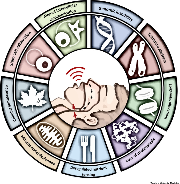
A heightened oxygen desaturation index was significantly linked to epigenetic age acceleration determined via two epigenetic clocks, according to a presentation at the American Thoracic Society International Conference.
“OSA severity is associated with positive epigenetic age acceleration or premature aging,” Ilia Ostrovski, respirology fellow from University of British Columbia, said during his presentation. “We also conclude that the association between obstructive sleep apnea and positive epigenetic age acceleration is better demonstrated by second generation epigenetic clocks likely due to their calibration with physiologic outcomes rather than chronological age.”
In a large, cross-sectional study, Ostrovski and colleagues evaluated epigenetic age, a biologic age biomarker, of 1,254 adults (mean age, 53 years; 43% women) from the 2016 to 2019 Canadian Sleep and Circadian Network biobank who underwent sleep testing to find out if OSA severity is linked to premature aging, or epigenetic age acceleration.
Researchers utilized blood samples to extract DNA and four validated epigenetic clocks to find epigenetic age estimations.
“Levels of DNA methylation at certain sites in the genome are associated with chronological age; thus, epigenetic or biological age can be predicted exploiting this feature of the DNA methylome,” Ostrovski and colleagues wrote in the study abstract.
Ostrovski noted during his presentation that first generation clocks (Horvath pan-tissue and Hannum) are calibrated to chronologic age, whereas second generation clocks (PhenoAge and GrimAge) are calibrated to physiologic outcomes and mortality.
“It’s becoming increasingly recognized that these [second generation clocks] capture age-related decline better than first generation clocks,” he said.
The total cohort included 325 controls (median age, 54 years; 54% women; median BMI, 28 kg/m2) and 929 individuals with OSA.
Of those with OSA, most had severe OSA (n = 387; median age, 56 years; 34% women; median BMI, 37 kg/m2), followed by moderate OSA (n = 297; median age, 56 years; 36% women; median BMI, 33 kg/m2) and mild OSA (n = 245; median age, 58 years; 51% women; median BMI, 31 kg/m2).
Baseline characteristics with a greater proportion of those with severe or moderate OSA vs. those with mild OSA or controls included current smoking status (14% vs. 12% vs. 9% vs. 7.4%), diabetes (28% vs. 18% vs. 17% vs. 9.3%), hypertension (51% vs. 55% vs. 42% vs. 32%) and cardiovascular disease (14% vs. 15% vs. 13% vs. 9%).
The proportion of individuals with alcohol use disorder was similar among controls, those with mild OSA and those with moderate OSA (63% vs. 62% vs. 63%) but lower among those with severe OSA (52%).
Further, median oxygen desaturation index (ODI) at baseline increased as OSA became more severe, starting at two desaturation episodes per hour in the control group and escalating to 10 episodes per hour in the mild OSA group, 21 episodes per hour in the moderate group and 50 episodes per hour in the severe group.
To find the relationship between epigenetic age acceleration and ODI for each clock, researchers used linear regression adjusted for blood cell type proportions, age, sex, ethnicity, smoking, alcohol use, BMI, chip ID and chip position/row.
Using the GrimAge clock, each rise in ODI by 10 corresponded to epigenetic age acceleration of 0.16 years (P = .004). The same increase in ODI was also linked to epigenetic age acceleration of 0.11 years in the PhenoAge clock (P = .039).
No significant relationship was found between the two factors when using each of the first-generation clocks, and Ostrovski highlighted that the link became nonsignificant after factoring in BMI.
“We feel that future work should prioritize prospective evaluation of the impact of OSA severity on aging-related outcomes and also determine whether treatment of OSA reverses its effect on epigenetic age acceleration,” Ostrovski said.
Professor, Department of Psychiatry Director, Center for Novel Therapeutics University of Colorado School of Medicine Anschutz Medical Campus
Researchers from the University of Colorado Anschutz Medical Campus have established a new framework for understanding how classic antidepressants work in treating major depressive disorder (MDD), reemphasizing their importance and aiming to reframe clinical conversation around their role in treatment.
The nature of the dysfunction at the root of MDD has been under investigation for decades. Classic antidepressants, like SSRIs (selective serotonin reuptake inhibitors, such as Prozac) cause an elevation in the levels of the brain chemical messenger, serotonin. This observation led to the idea that antidepressants work because they restore a chemical imbalance, such as a lack of serotonin. However, subsequent years of research showed no significant decrease in serotonin in people with depression. While experts have moved away from this hypothesis due to lack of concrete evidence, this has led to a shift in public opinion on the effectiveness of these medications.
Antidepressants, such as SSRIs and serotonin and norepinephrine reuptake inhibitors (SNRIs) are still effective in alleviating depressive episodes in many patients, however. In a paper published in Molecular Psychiatry, researchers outline a new framework for understanding how antidepressants are efficacious in treating MDD. This framework helps clarify how antidepressants like SSRIs are still be helpful, even if MDD isn’t caused by a lack of serotonin.
“The best evidence of changes in the brain in people suffering from MDD is that some brain regions are not communicating with each other normally,” says Scott Thompson, PhD, professor in the department of psychiatry at the University of Colorado School of Medicine and senior author. “When the parts of the brain responsible for reward, happiness, mood, self-esteem, even problem solving in some cases, are not communicating with each other properly, then they can’t do their jobs properly.
“There is good evidence that antidepressants that increase serotonin, like SSRIs, all work by restoring the strength of the connections between these regions of the brain. So do novel therapeutics such as esketamine and psychedelics. This form of neuroplasticity helps release brain circuits from being ‘stuck’ in a pathological state, ultimately leading to a restoration of healthy brain function,” said Thompson.
Thompson and colleagues liken this theory to a car running off the road and getting stuck in a ditch, requiring the help of a tow truck to pull the car out of its stuck state, allowing it to move freely down the road again.
Researchers are hoping health care providers will use their examples to bolster conversations with apprehensive patients about these treatments, helping them better understand their condition and how to treat it.
“We are hoping this framework provides clinicians new ways to communicate the way these treatments work in combating MDD,” said C. Neill Epperson, MD, Robert Freedman endowed professor and chair of the department of psychiatry in the University of Colorado School of Medicine and co-author on the paper. “Much of the public conversation around the effectiveness of antidepressants, and the role serotonin plays in diagnosis and treatment, has been negative and largely dangerous. While MDD is a heterogenous disorder with no one fits all solution, it is important to emphasize that if a treatment or medication is working for you, then they are lifesaving. Understanding how these medications promote neuroplasticity can help strengthen that message.”
Chloe E. Page, C. Neill Epperson, Andrew M. Novick, Korrina A. Duffy, Scott M. Thompson. Beyond the serotonin deficit hypothesis: communicating a neuroplasticity framework of major depressive disorder. Molecular Psychiatry, 2024; DOI: 10.1038/s41380-024-02625-2
BERLIN (AP) — When Michael Bommer found out that he was terminally ill with colon cancer, he spent a lot of time with his wife, Anett, talking about what would happen after his death.
She told him one of the things she’d miss most is being able to ask him questions whenever she wants because he is so well read and always shares his wisdom, Bommer recalled during a recent interview with The Associated Press at his home in a leafy Berlin suburb.
That conversation sparked an idea for Bommer: Recreate his voice using artificial intelligence to survive him after he passed away.
The 61-year-old startup entrepreneur teamed up with his friend in the U.S., Robert LoCascio, CEO of the AI-powered legacy platform Eternos. Within two months, they built “a comprehensive, interactive AI version” of Bommer — the company’s first such client.
Eternos, which got its name from the Italian and Latin word for “eternal,” says its technology will allow Bommer’s family “to engage with his life experiences and insights.” It is among several companies that have emerged in the last few years in what’s become a growing space for grief-related AI technology.
One of the most well-known start-ups in this area, California-based StoryFile, allows people to interact with pre-recorded videos and uses its algorithms to detect the most relevant answers to questions posed by users. Another company, called HereAfter AI, offers similar interactions through a “Life Story Avatar” that users can create by answering prompts or sharing their own personal stories.
There’s also “Project December,” a chatbot that directs users to fill out a questionnaire answering key facts about a person and their traits — and then pay $10 to simulate a text-based conversation with the character. Yet another company, Seance AI, offers fictionalized seances for free. Extra features, such as AI-generated voice recreations of their loved ones, are available for a $10 fee.
While some have embraced this technology as a way to cope with grief, others feel uneasy about companies using artificial intelligence to try to maintain interactions with those who have passed away. Still others worry it could make the mourning process more difficult because there isn’t any closure.
Katarzyna Nowaczyk-Basinska, a research fellow at the University of Cambridge’s Centre for the Future of Intelligence who co-authored a study on the topic, said there is very little known about the potential short-term and long-term consequences of using digital simulations for the dead on a large scale. So for now, it remains “a vast techno-cultural experiment.”
“What truly sets this era apart — and is even unprecedented in the long history of humanity’s quest for immortality — is that, for the first time, the processes of caring for the dead and immortalization practices are fully integrated into the capitalist market,” Nowaczyk-Basinska said.
Bommer, who only has a few more weeks to live, rejects the notion that creating his chatbot was driven by an urge to become immortal. He notes that if he had written a memoir that everyone could read, it would have made him much more immortal than the AI version of himself.
“In a few weeks, I’ll be gone, on the other side — nobody knows what to expect there,” he said with a calm voice.
PRESERVING A CONNECTION
Robert Scott, who lives in Raleigh, North Carolina, uses AI companion apps Paradot and Chai AI to simulate conversations with characters he created to imitate three of his daughters. He declined to speak about what led to the death of his oldest daughter in detail, but he lost another daughter through a miscarriage and a third who died shortly after her birth.
Scott, 48, knows the characters he’s interacting with are not his daughters, but he says it helps with the grief to some degree. He logs into the apps three or four times a week, sometimes asking the AI character questions like “how was school?” or inquiring if it wants to “go get ice cream.”
Some events, like prom night, can be particularly heart-wrenching, bringing with it memories of what his eldest daughter never experienced. So, he creates a scenario in the Paradot app where the AI character goes to prom and talks to him about the fictional event. Then there are even more difficult days, like his daughter’s recent birthday, when he opened the app and poured out his grief about how much he misses her. He felt like the AI understood.
“It definitely helps with the what ifs,” Scott said. “Very rarely has it made the ‘what if’s’ worse.”
Matthias Meitzler, a sociologist from Tuebingen University, said that while some may be taken aback or even scared by the technology — “as if the voice from the afterlife is sounding again” — others will perceive it as an addition to traditional ways of remembering dead loved ones, such as visiting the grave, holding inner monologues with the deceased, or looking at pictures and old letters.
But Tomasz Hollanek, who worked alongside Nowaczyk-Basinska at Cambridge on their study of “deadbots” and “griefbots,” says the technology raises important questions about the rights, dignities and consenting power of people who are no longer alive. It also poses ethical concerns about whether a program that caters to the bereaved should be advertising other products on its platform, for example.
“These are very complicated questions,” Hollanek said. “And we don’t have good answers yet.”
Another question is whether companies should offer meaningful goodbyes for someone who wants to cease using a chatbot of a dead loved one. Or what happens when the companies themselves cease to exist? StoryFile, for example, recently filed for Chapter 11 bankruptcy protection, saying it owes roughly $4.5 million to creditors. Currently, the company is reorganizing and setting up a “fail-safe” system that allows families to have access to all the materials in case it folds, said StoryFile CEO James Fong, who also expressed optimism about its future.
PREPARING FOR DEATH
The AI version of Bommer that was created by Eternos uses an in-house model as well as external large language models developed by major tech companies like Meta, OpenAI and the French firm Mistral AI, said the company’s CEO LoCascio, who previously worked with Bommer at a software company called LivePerson.
Eternos records users speaking 300 phrases — such as “I love you” or “the door is open” — and then compresses that information through a two-day computing process that captures a person’s voice. Users can further train the AI system by answering questions about their lives, political views or various aspects of their personalities.
The AI voice, which costs $15,000 to set up, can answer questions and tell stories about a person’s life without regurgitating pre-recorded answers. The legal rights for the AI belongs to the person on whom it was trained and can be treated like an asset and passed down to other family members, LoCascio said. The tech companies “can’t get their hands on it.”
Because time has been running out for Bommer, he has been feeding the AI phrases and sentences — all in German — “to give the AI the opportunity not only to synthesize my voice in flat mode, but also to capture emotions and moods in the voice.” And indeed the AI voicebot has some resemblance with Bommer’s voice, although it leaves out the “hmms” and “ehs” and mid-sentence pauses of his natural cadence.
Sitting on a sofa with a tablet and a microphone attached to a laptop on a little desk next to him and pain killer being fed into his body by an intravenous drip, Bommer opened the newly created software and pretended being his wife, to show how it works.
He asked his AI voicebot if he remembered their first date 12 years ago.
“Yes, I remember it very, very well,” the voice inside the computer answered. “We met online and I really wanted to get to know you. I had the feeling that you would suit me very well — in the end, that was 100% confirmed.”
Bommer is excited about his AI personality and says it will only be a matter of time until the AI voice will sound more human-like and even more like himself. Down the road, he imagines that there will also be an avatar of himself and that one day his family members can go meet him inside a virtual room.
In the case of his 61-year-old wife, he doesn’t think it would hamper her coping with loss.
“Think of it sitting somewhere in a drawer, if you need it, you can take it out, if you don’t need it, just keep it there,” he told her as she came to sit down next to him on the sofa.
But Anett Bommer herself is more hesitant about the new software and whether she’ll use it after her husband’s death.
Right now, she more likely imagines herself sitting on the couch sofa with a glass of wine, cuddling one of her husband’s old sweaters and remembering him instead of feeling the urge to talk to him via the AI voicebot — at least not during the first period of mourning.
“But then again, who knows what it will be like when he’s no longer around,” she said, taking her husband’s hand and giving him a glance.
Grieshaber is a Berlin-based reporter covering Germany and Austria for The Associated Press. She covers general news as well as migration, populism and religion.
Increased risk for autism spectrum disorder was associated with 7.36 µg/L lithium exposure.
Increased risk for schizophrenia spectrum disorder was associated with 5.8 µg/L lithium exposure.
NEW YORK — Lithium exposure in drinking water was associated with potentially detrimental effects on human health, including increased risk for autism spectrum disorder and schizophrenia spectrum disorder, according to researchers.
“At the state hospital we deal with a lot of the sickest patients,” Sonja M. Johnson, DO, a fourth-year psychiatry resident at Indiana University Health, told Healio at the American Psychiatric Association annual meeting. “Lithium is an awesome medication, and it does a lot of great things. We always hear the phrase, ‘That’s so good. Put it in the water!’ I mean, they did it with fluoride, right?”
Andrea Patterson, MD, also a fourth-year resident in psychiatry, and colleagues at Indiana University School of Medicine performed a systematic review of 26 studies with data from five continents to determine whether higher levels of environmental lithium in the water supply poses a risk to human health.
Sonja M. Johnson
“Mental health is still kind of taboo in our area, but water is important, and people drink water,” Johnson said. “The question was, can we help everyone without making everyone take medicine?”
Of the reviewed studies, 12 showed that lithium exposure through drinking water had the potential for negative effects on the nervous, cardiovascular, endocrine, lymphatic, urinary and integumentary systems and could affect newborns to adults and pregnant women.
Although researchers reported that at 7 µg/L, lithium begins to have protective factors against suicide, they noted that at 7.36 µg/L it was associated with autism spectrum disorder, and at 5.8 µg/L with schizophrenia.
“Given this information, any lithium added to the U.S. water supply for protective reasons would inevitably increase the risk of harm,” researchers wrote.
“When you ask a question, sometimes the answer is no,” Johnson said. “That’s still an answer, and that’s still pretty awesome, because if you don’t ask you don’t know.”
Patterson A, et al. Too much of a good thing? Detrimental health effects linked to environmental lithium exposure through drinking water: A systematic review. Presented at: American Psychiatric Association annual meeting; May 4-8, 2024; New York.
Disclosures: Johnson reported no relevant financial disclosures. Please see the study for all other authors’ relevant financial disclosures.