In a groundbreaking experiment, scientists have successfully transmitted both quantum and conventional internet data through the same fiber-optic channel.This significant achievement was documented in a study published in the journal Science Advances.The research indicates that quantum data, represented by entangled photons, can coexist with traditional internet data transmitted as laser pulses within a single fiber-optic cable.
Quantum leap
A breakthrough in quantum communication
The successful transmission of both quantum and conventional data through a single optical fiber marks a significant advancement in the field of quantum communication.This achievement challenges previous research that suggested separate infrastructure or dedicated channels were required for quantum data to avoid interference from “classical” data.The new “hybrid” network could possibly allow for more efficient implementation of quantum communications by enabling both types of data to share the same infrastructure.
Technical hurdles
Overcoming challenges in creating hybrid networks
Creating hybrid networks is a complex task due to the delicate nature of quantum data transmission via fiber-optic cables using entangled photons.Entanglement, a state where two qubits share information irrespective of their relationship over time or space, can be easily disrupted by environmental disturbances such as noise or interference from other signals.This disruption, also known as “decoherence,” causes the qubits to lose their quantum state and results in data loss.
To overcome the challenges of creating hybrid networks, scientists used an innovative technique called electro-optic phase modulation.This method permitted them to precisely adjust the frequency of laser pulses to match the color of entangled photons.As a result, both types of data could be transmitted in the same color channel without disrupting quantum information held by entangled photons.
Quantum potential
It could revolutionize quantum computing applications
The successful transmission of both quantum and conventional data in the same channel could potentially free up other color channels in the fiber-optic cable for more data.This development is crucial for making applications of quantum computing, like quantum cryptography and ultra-secure communications, more practical and scalable.”Our research is an important step to combine the conventional internet with the quantum internet,” said study co-author Michael Kues.
Those who sustained a concussion on artificial turf were older than those who sustained a concussion on grass.
The groups were similar in number of days before evaluation, concussion and headache history.
In young male American football players, concussion symptoms and severity were higher for those who sustained the injury on natural grass compared with artificial turf, according to new research in the Clinical Journal of Sports Medicine.
“Many natural grass fields, especially at the youth level, may not be well-maintained and can be harder and less forgiving than modern artificial turf, which has evolved significantly from the old, hard fields of the past,” C. Munro Cullum, PhD, lead study author and professor of clinical psychiatry in the Peter J, O’Donnell Brain Institute at the University of Texas Southwestern Medical Center, said in a related release.
Cullum and colleagues examined the differences in concussion symptoms for young American football players who were injured on natural grass and artificial surfaces.
Their study culled data from the prospective, longitudinal, multi-institutional North Texas Concussion Registry (ConTex) research project to include 62 male football players aged 10 to 24 years who sustained a helmet-to-ground impact concussion (grass impact, n = 33; artificial turf impact, n = 29) and presented to a specialty concussion clinic within 14 days of the injury.
The main outcome for analysis was self-reported, post-injury number and severity of symptoms measured by the 22-item Sport Concussion Assessment Tool 5th Edition (SCAT5) Symptom Evaluation at the time of participants’ first clinic visit to UT Southwestern Medical Center. SCAT5 scores are combined on a scale ranging from 0 (no symptoms) to six (most severe).
According to the results, both groups had a similar mean number of days since injury before evaluation (6.1 vs. 5.3 days), concussion history and headache history.
However, the researchers wrote that players who sustained a concussion from contact on a grass-playing field reported higher mean total symptom severity scores (26.6 vs. 11.6, P = .005) and total number of symptoms (10.3 vs. 5.9, P = .006) compared with those who were injured on artificial turf.
Data additionally showed those in the artificial turf group were slightly older than those in the natural grass cohort (mean age, 14.6 years vs. 13.6 years).
Cullum and colleagues noted key limitations to their work, chief among them a small sample size as well as a significant mean age difference between the two groups, suggesting that either older athletes are more likely to play on artificial surfaces, or that the result is due to more playing experience.
The findings conflict with previous research that suggest young athletes may be at greater risk for head injuries on synthetic turf. However, several other studies have found “lower rates of football-related concussion on artificial turf versus grass,” Cullum said in the release.
“Our study also suggests that concussions on natural surfaces are more likely to be worse and may require longer recovery times,” he said.
These ‘living computers’ are made from human neurons — and you can rent one for $500 a month.
By Jordan Kinard
Artificial intelligence systems, even those as sophisticated as ChatGPT, depend on the same silicon-based hardware that has been the bedrock of computing since the 1950s. But what if computers could be molded from living biological matter? Some researchers in academia and the commercial sector, wary of AI’s ballooning demands for data storage and energy, are focusing on a growing field known as biocomputing. This approach uses synthetic biology, such as miniature clusters of lab-grown cells called organoids, to create computer architecture. Biocomputing pioneers include Swiss company FinalSpark, which earlier this year debuted its “Neuroplatform”—a computer platform powered by human-brain organoids—that scientists can rent over the Internet for $500 a month.
“As far as I know, we are the only ones in the world doing this” on a publicly rentable platform, says FinalSpark co-founder Fred Jordan. Initially bankrolled with funds from its co-founders’ previous start-up, FinalSpark seeks an environmentally sustainable way to support AI. “Our principal goal is artificial intelligence for 100,000 times less energy” than what’s currently required to train state-of-the-art generative AI, Jordan says. Neuroplatform uses a series of processing units hosting four spherical brain organoids each. Every 0.5-millimeter-wide organoid is connected to eight electrodes that electrically stimulate the neurons within the living sphere; those electrodes also link the organoids to conventional computer networks. The neurons are selectively exposed to the feel-good neurotransmitter dopamine to mimic the human brain’s natural reward system. These twin setups—positive dopamine rewards and electrical stimulation—train the organoids’ neurons, prompting them to form new pathways and connections much in the same way a living human brain appears to learn. If perfected, this training could eventually allow organoids to mimic silicon-based AI and serve as processing units with functions similar to today’s CPUs (central processing units) and GPUs (graphics processing units), FinalSpark says.
For now, the organoids and their behavior are live streamed 24 hours a day for researchers (and anyone else) to observe. “The challenge is to find the appropriate way to get neurons to do what we want them to do,” Jordan says.
Research teams at 34 universities have asked to use FinalSpark’s biocomputers, and so far the company has provided access for scientists at the University of Michigan, the Free University of Berlin and seven other institutions. Each one’s project focuses on a different aspect of biocomputing. The University of Michigan team, for example, is investigating the electrical and chemical prompts necessary to change organoid activity—in effect creating the building blocks of an organoid-specific computer language. Scientists at Lancaster University Leipzig in Germany, meanwhile, are trying to fit the organoids into different models of AI learning.
Sticking points remain for organoid computing’s ability to compete with silicon on a large scale. For one thing, no standardized manufacturing system exists. And living brains die: FinalSpark’s organoids only survive for an average of around 100 days (and that’s considerable progress from the original experiment’s lifespan, which was just a few hours). But Jordan notes that Neuroplatform has “streamlined” its in-house process for making organoids, and its facility currently houses between 2,000 and 3,000 of them.
FinalSpark is not alone in its pursuit of organic alternatives to silicon-based computing, and brain organoids are not the only possible way forward. “There are different flavors of biocomputing,” says Ángel Goñi-Moreno, a researcher at Spain’s National Center for Biotechnology. Goñi-Moreno studies cellular computing, or the use of modified living cells to create systems that can replicate “memory, logic gates and the other decision-making basics we know from conventional computer science,” he says. His team is looking for tasks at which biocomputers outperform their silicon counterparts—a dynamic he calls “cellular supremacy.” In particular, Goñi-Moreno believes that because cellular computers can react to their environmental conditions, they could facilitate bioremediation, or the restoration of damaged ecosystems. “That’s a domain where conventional computers can do basically nothing,” Goñi-Moreno says. “You can’t just throw a computer into a lake and have it tell you the state of the environment.” A submerged bacterial computer, however, would be able to give a nuanced reading of environmental conditions as the cells respond to chemical and other stimuli.
Where Goñi-Moreno is focused on bacteria, Andrew Adamatzky of the University of the West of England, founding editor in chief of the International Journal of Unconventional Computing, has been studying the computational possibilities of fungus. Mycelia, or networks of fungal strands, exhibit spiking electrical potentials similar to those found in neurons, Adamatzky says. He hopes to take advantage of these electrical properties to create a brainlike fungal computing system that is “potentially capable of learning, reservoir computing, pattern recognition, and more.” Adamatzky’s team has already successfully trained fungal networks to help computer systems perform certain mathematical functions. “Fungal computing offers several advantages over brain-organoid-based computing,” Adamatzky says, “particularly in terms of ethical simplicity, ease of cultivation, environmental resilience, cost-effectiveness and integration with existing technologies.”
Jordan is well aware of the considerations involved in using cultivated human neurons for nonmedical purposes. An ongoing bioethical debate concerns whether mini brains could gain consciousness, though there is as yet no evidence it has ever been created in a lab. Jordan says he is currently seeking philosophers and researchers with the “cultural background to help us answer these ethical questions.”
Adamatzky acknowledges that brain organoids “might offer advanced functionalities due to their complex and neuronlike structures” despite his advocacy of fungal computing. Jordan, for his part, is confident in FinalSpark’s choice for its biocomputers. Of all the cells to pick from, he says, “human neurons are the best at learning.”
Earth’s days were once more than two hours longer than they are now, thanks to the moon drifting thousands of miles farther away in its orbit over two periods, researchers have discovered.
The extra hours of sunlight, in turn, may have led to oxygenation events that ushered in a period when life’s complexity exploded, the study researchers say.
“Daylength changes may influence the distribution of solar energy and temperature gradients, potentially impacting weather systems and atmospheric dynamics,” the researchers wrote in the new study, published Aug. 6 in the journal PNAS.
Earth’s days are currently around 24 hours long, but that wasn’t always the case. Over time, the moon tugs on our planet. As it does so, it migrates away from Earth, siphoning away its kinetic energy. As a result, our planet’s spin around its axis slows, thus lengthening Earth’s days, according to the study.
Modeling changes in how Earth wobbles as it spins can give a rough picture of this slowdown over the planet’s history. But this estimate is clearly flawed, because it leads to a prediction that Earth and the moon would have collided around 1.5 billion years ago, the study authors noted.
In the new study, a team led by geologist He Huang from the Chengdu University of Technology in China, tried to clarify Earth’s spin history by looking at eight datasets that captured rock layers from marine environments dating to roughly between 700 million and 200 million years ago. These tidalites, as they’re called, can record the strength of the tides over time, in part because they reveal the ocean’s thickness. The team combined these datasets with models of the tidal forces acting between the moon and Earth to map how quickly Earth spun around its axis over the half-billion-year study period.
The researchers found that there was a “staircase” pattern in Earth’s spin, with two periods where the planet’s rotation quickly and dramatically changed, followed by periods of stability. Over the study period, days were 2.2 hours longer than they are now. The moon, during this period, was also an average of 12,000 miles (20,000 km) farther away.
One of these time periods, roughly 650 million to 500 million years ago, encompassed the Cambrian explosion, a period when life diversified dramatically and radiated into new niches. The second “step” in the staircase of Earth’s spin occurred roughly 340 million to 280 million years ago, which corresponded to a period when massive glaciers covered the planet.
The study suggests that by increasing the day length — and, therefore, sun exposure — the moon may have triggered great oxygenation events that led to life’s diversification. However, those results “need to be interpreted with care,” the authors wrote in the study.
The study also casts doubt on another theory: that a period when glaciers rapidly covered Earth, leading to a mass extinction, had a big effect on Earth’s spin. Rather, most of the slowdown is caused directly by tidal forces, they suggested.
In a study, published in the journal The Proceedings of the National Academy of Sciences, a molecule identified and synthesized by UCLA Health researchers was shown to restore cognitive functions in mice with symptoms of Alzheimer’s disease by effectively jump-starting the brain’s memory circuitry.
If proven to have similar effects in humans, the candidate compound would be novel among Alzheimer’s disease treatments in its ability to revitalize memory and cognition, the study authors said.
“There is really nothing like this on the market or experimentally that has been shown to do this,” said study lead author Dr. Istvan Mody, a professor of neurology and physiology at UCLA Health.
The molecule, DDL-920, works differently from recent FDA-approved drugs for Alzheimer’s disease such as lecanemab and aducanumab, which remove harmful plaque that accumulates in the brains of Alzheimer’s disease patients. While removing this plaque has been shown to slow the rate of cognitive decline, it does not restore memory and cognitive impairments.
“They leave behind a brain that is maybe plaqueless, but all the pathological alterations in the circuits and the mechanisms in the neurons are not corrected,” Mody said.
In the study, titled “A therapeutic small molecule enhances γ-oscillations and improves cognition/memory in Alzheimer’s disease model mice,” UCLA researchers led by Dr. Istvan Mody and Dr. Varghese John, professor of neurology, and director of the Drug Discovery Laboratory (DDL) at the Mary S. Easton Center for Alzheimer’s Disease Research and Care sought to find a compound that could figuratively flip the switch back on in the brain’s memory circuitry.
Similar to a traffic signal, the brain fires off electric signals at different rhythms to start and stop various functions. Gamma oscillations are some of the highest-frequency rhythms and have been shown to orchestrate brain circuits underlying cognitive processes and working memory—the type of memory used to remember a phone number.
Patients with early Alzheimer’s disease symptoms such as mild cognitive impairment have been shown to have reduced gamma oscillations, Mody said.
Other studies attempted to use neuromodulation techniques to stimulate gamma oscillations to restore memory. Auditory, visual or transcranial magnetic stimulation at a frequency of 40 Hz—similar to the frequency of a cat’s purr—worked to dissolve plaques in the brain but again did not show notable cognitive enhancements, Mody said.
In this latest study, Mody and his team sought to tackle the problem from a different perspective. If they could not jump-start these memory circuits using external tools, perhaps there was a way to trigger these electrical rhythms from the inside using a molecule.
Specifically, they needed a compound to target certain fast-firing neurons, known as the paravalbumin interneurons, that are critical in generating gamma oscillations and therefore memory and cognitive functions. However, certain chemical receptors in these neurons that respond to the chemical messenger known as GABA work like brake pedals to reduce the gamma oscillations entrained by these neurons.
Mody, John and their team identified the compound DDL-920 to antagonize these receptors, allowing the neurons to sustain more powerful gamma oscillations.
To test whether this would actually result in improved memory and cognition, researchers used mice that were genetically modified to have symptoms of Alzheimer’s disease.
Both these Alzheimer’s disease model mice and wild-type mice underwent baseline cognitive testing in a Barnes maze—a circular platform surrounded by visual clues and containing one escape hole. The maze is used to measure how well rodents can learn and remember the location of the escape hole.
After the initial tests, researchers orally administered DDL-920 to the Alzheimer’s model mice twice daily for two weeks. Following treatment, the Alzheimer’s disease model mice were able to recall the escape hole in the maze at similar rates as the wild-type mice. Additionally, the treated mice did not display any abnormal behavior, hyperactivity or other visible side effects over the two-week period.
Mody said that while the treatment was effective in mice, much more work would be needed to determine if the treatment would be safe and effective in humans. Should it ultimately prove to be effective, the drug could have implications for treatments of other diseases and health conditions that have diminished gamma oscillations such as depression, schizophrenia and autism spectrum disorder, Mody said.
“We are very enthusiastic about that because of the novelty and the mechanism of action that has not been tackled in the past,” Mody said.
More information: Xiaofei Wei et al, A therapeutic small molecule enhances γ-oscillations and improves cognition/memory in Alzheimer’s disease model mice, Proceedings of the National Academy of Sciences (2024). DOI: 10.1073/pnas.2400420121
Whether Bach, Beethoven, or Mozart, it’s widely recognized that classical music can affect a person’s mood. In a study published in Cell Reports, scientists in China have used brainwave measurements and neural imaging techniques to show how Western classical music elicits its positive effects on the brain. Their goal is to find more effective ways to use music to activate the brain in those who otherwise don’t respond, such as people with treatment-resistant depression.
“Our research integrates the fields of neuroscience, psychiatry, and neurosurgery, providing a foundation for any research targeting the interaction between music and emotion,” says senior author Bomin Sun, director and professor of the Center for Functional Neurosurgery at Shanghai Jiao Tong University. “Ultimately, we hope to translate our research findings into clinical practice, developing convenient and effective music therapy tools and applications.”
The study focused on 13 patients with treatment-resistant depression who already had electrodes implanted in their brains for the purpose of deep-brain stimulation. These implants are placed in a circuit connecting two areas in the forebrain—the bed nucleus of the stria terminalis (BNST) and the nucleus accumbens (NAc). Using these implants, the researchers found that music generates its antidepressant effects by synchronizing the neural oscillations between the auditory cortex, which is responsible for processing of sensory information, and the rewards circuit, which is responsible for processing emotional information.
“The BNST-NAc circuit, sometimes referred to as part of the ‘extended amygdala,’ underscores the close relationship between this circuit and the amygdala, a central structure in emotional information processing,” Sun says. “This study reveals that music induces triple-time locking of neural oscillations in the cortical-BNST-NAc circuit through auditory synchronization.”
The patients in the study were assigned to two groups: low music appreciation or high music appreciation. Those in the high music appreciation group demonstrated more significant neural synchronization and better antidepressant effects, while those in the low music appreciation group showed poorer results. By grouping the patients, the investigators were able to study the antidepressant mechanisms of music more precisely and propose personalized music therapy plans that would improve treatment outcomes. For example, when inserting theta frequency noise into music to enhance BNST-NAc oscillatory coupling, those in the low music appreciation group of patients reported higher music enjoyment.
Several pieces of Western classical music were used in the study. This type of music was chosen because most participants did not have familiarity with it, and the researchers wanted to avoid any interference that could arise from subjective familiarity.
“We concluded that the music choices during the formal listening process were individualized and unrelated to the music’s emotional background,” Sun says.
The team’s future research will focus on several areas. For one, they aim to study how the interaction between music and the deep structures of the brain play a role in depressive disorders. They will also introduce other forms of sensory stimuli, including visual images, to investigate potential combined therapeutic effects of multi-sensory stimulation on depression.
“By collaborating with clinicians, music therapists, computer scientists, and engineers, we plan to develop a series of digital health products based on music therapy, such as smartphone applications and wearable devices,” Sun says. “These products will integrate personalized music recommendations, real-time emotional monitoring and feedback, and virtual-reality multi-sensory experiences to provide convenient and effective self-help tools for managing emotions and improving symptoms in daily life.”
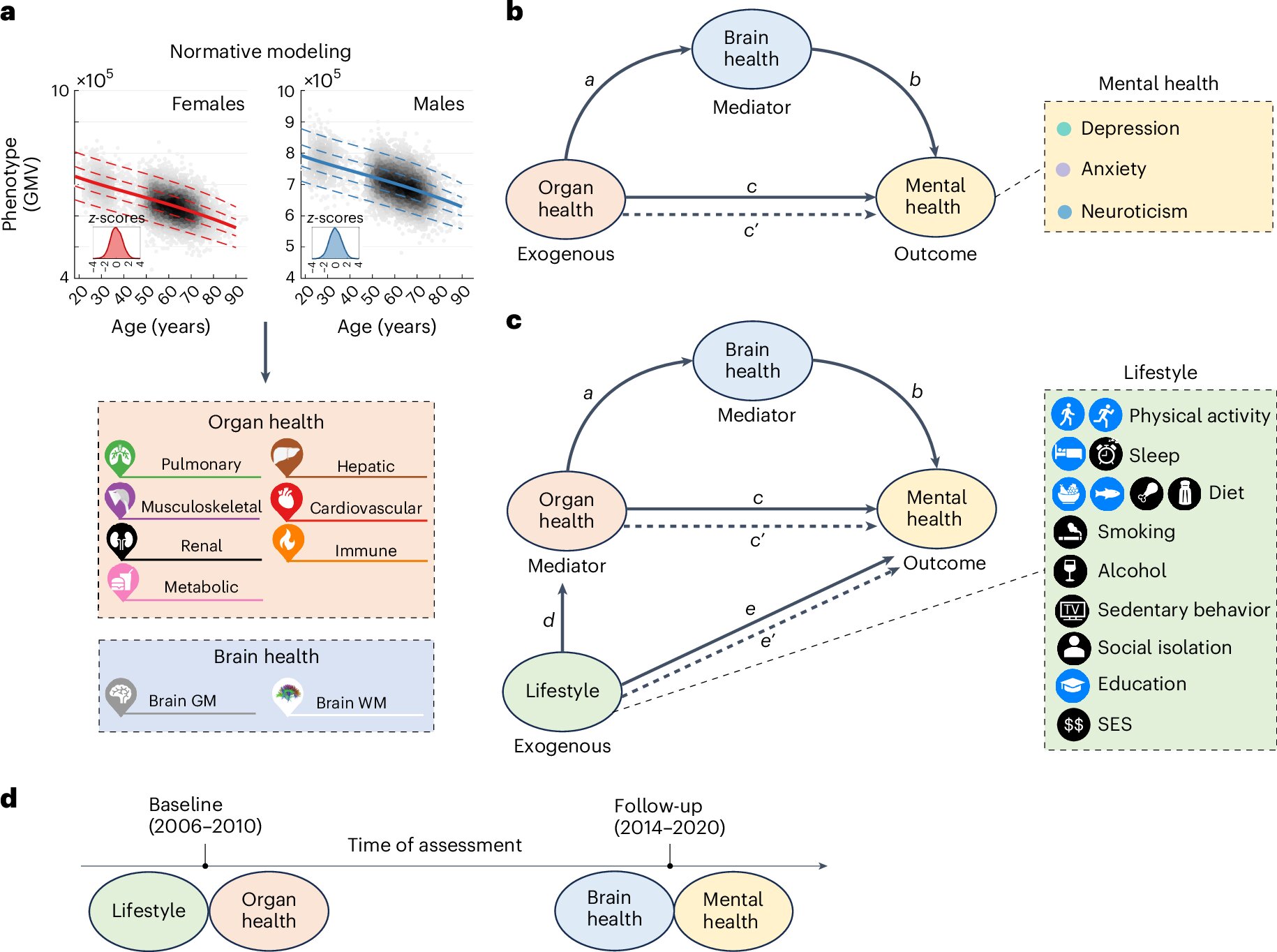
The interconnectedness of the brain, body and lifestyle factors and how they collectively influence mental health has been demonstrated by new research.
Researchers from the University of Melbourne, University College London and the University of Cambridge have identified multiple biological pathways involving organs and the brain that play a key part in physical and mental health.
The study, published today in Nature Mental Health, used UK Biobank data from more than 18,000 people—7,749 people in the study had no major clinically diagnosed medical or mental health conditions, while 10,334 reported a diagnosis of either schizophrenia, bipolar disorder, depression or anxiety.
Using advanced statistical models, the researchers found poorer organ health was significantly associated with higher depressive symptoms, and that the brain plays an important role in linking body health and depression.
The organ systems studied included the lungs, muscles and bones, kidneys, liver, heart, and the metabolic and immune systems.
“Overall, we found multiple significant pathways through which poor organ health may lead to poor brain health, which may in turn lead to poor mental health,” lead author Dr. Ye Ella Tian, research fellow in the Department of Psychiatry, said.
“By integrating clinical data, brain imaging and a wide array of organ-specific biomarkers in a large population-based cohort, we were able to establish for the first time multiple pathways involving the brain as a mediating factor and through which poor physical health of body organ systems may lead to poor mental health.
“We identified modifiable lifestyle factors that can potentially lead to improved mental health through their impact on these specific organ systems and neurobiology.
“Our work provides a holistic characterization of brain, body, lifestyle and mental health.”
Physical health was also taken into account as well as lifestyle factors such as sleep quality, diet, exercise, smoking, and alcohol consumption.
“This is a significant body of work because we have shown the link between physical health and depression and anxiety and how that is partially influenced by individual changes in brain structure,” Professor Andrew Zalesky from the Departments of Psychiatry and Biomedical Engineering said.
“Our results suggest that poor physical health across multiple organ systems, such as liver and heart, the immune system and muscles and bones, may lead to subsequent alterations in brain structure.
“These structural changes of the brain may lead to or exacerbate symptoms of depression and anxiety as well as neuroticism.”
Professor James Cole, an author of the study from UCL Computer Science, said, “While it’s well-known in health care that all the body’s organs and systems influence each other, it’s rarely reflected in research studies. So, it’s exciting to see these results, as it really emphases the value in combining measures from different parts of the body together.”
More information: Ye Ella Tian et al, Brain, lifestyle and environmental pathways linking physical and mental health, Nature Mental Health (2024). DOI: 10.1038/s44220-024-00303-4
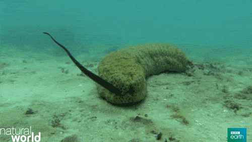
This slimline, eel-like fish has no scales for protection so chooses to use a sea cucumber’s sphincter for safety.
Name: Pearlfish (Carapidae family)
Where it lives: Inside invertebrate hosts in shallow, tropical waters around the world
What it eats: Plankton, small particles and sea cucumbers’ gonads
Pearlfish don’t have scales or any way of protecting themselves, so instead they have to find a safe hideaway. But rather than sheltering in a seagrass meadow or hiding in the crevices of a rock, they have chosen an unusual refuge: sea cucumbers’ anuses.
“The rear end of a sea cucumber may seem like undesirable real estate, but for a pearlfish, it does just the trick,” according to the Smithsonian Ocean website.
Sea cucumbers breathe through their butts, giving pearlfish an easy opportunity to sneak into their unwitting host. The pearlfish sniffs out its host, then just has to wait “for the cucumber to open for a breath and swim inside,” Smithsonian Ocean said. They do this every time they need to re-enter their home, which they leave to find food.
Even the sea cucumbers’ Cuvierian tubules — special sticky threads that sea cucumbers eject from their bums in self-defense — don’t affect pearlfish. And cucumber species with anal teeth still succumb to these pesky butt-dwellers. “I have found a new species that is smaller that was able to go inside, even with the teeth,” Eric Parmentier, a pearlfish researcher at the University of Liège, Belgium, told Live Science in an email.
These eel-like fish often — but not always — make a home for one inside their host. “As far as we know, there is usually one [pearlfish] to a sea cucumber, but some species have been reported to pair up in a single cucumber,” Matt Girard, a researcher in the Division of Fishes at the Smithsonian National Museum of Natural History told Live Science in an email.
The number of pearlfish shacked up in a single sea cucumber can hit double figures. In 1975, scientist Victor Benno Meyer-Rochow found a leopard sea cucumber (Bohadschia argus) with 15 pearlfish inside it.
Some species can live inside sea cucumbers without doing them harm, Girard said. They live in symbiosis and “neither host nor invader is harmed.”
But there are also parasite species that “eat the sea cucumber’s gonads,” Parmentier added. “It does not kill the host but it can disturb its reproduction.”
A cohort of healthy midlife adults were randomized to eat both low- and high-sugar diets for 10 days.
The high-sugar diet adversely affected both cognitive ability and cardiometabolic health.
PHILADELPHIA — Consuming a high-sugar diet over 10 days was associated with memory issues as well as negative cardiometabolic health compared with a low-sugar diet, according to a poster presentation.
“We’re interested in some of the lifestyle/behavioral factors, but modifiable risk factors, like diet, are aspects that are a little bit more controllable,” Kevin P. Decker, PhD, postdoctoral researcher in the department of kinesiology and applied physiology at the University of Delaware, told Healio at the Alzheimer’s Association International Conference.
Working with the hypothesis that higher sugar intake will have a deleterious effect on blood pressure, Decker and colleagues sought to determine if a spike in added sugars over the course of 10 days given to midlife adults could diminish memory recall and decrease cardiometabolic health.
Specifically, their study compared diets consisting of 2,000 calories per day in which sugar accounted for 20% to 25% of total calories vs. 5% of total calories in 38 healthy adults (63.2% female, mean age 57±4 years) with no prior history of cardiovascular disease.
The study was divided into four phases. In the initial phase, participants were required to keep a 3-day diet record so the researchers could determine eligibility. Once it was established, enrollees were given baseline tests, including BP and blood-based biomarkers, then randomly assigned to receive either diet. The tertiary phase was randomization, during which participants picked up the 10-day supply of food and drink to be consumed at their own leisure with a “meal plan” guideline. At the end of the 10 days, all submitted to further BP and biomarker testing, along with cognitive evaluations (Brief Visuospatial Memory Test [BVMT]; Hopkins Verbal Learning Test [HVLT]).
Following a minimum 2-week washout period to prevent carryover effects from initial randomization, the diets were flipped; those given a high-sugar diet were switched to low-sugar and vice versa.
According to the results, the 10-day, high-sugar diet led to lower total memory recall in the BVMT compared with the low-sugar alternative, while also leading to delayed memory recall in both the BVMT and HVLT.
The researchers further discovered the high-sugar diet negatively impacted cardiometabolic health, with elevated triglycerides and BP compared with the low-sugar diet.
“We saw that there was lower memory recall on the high-sugar diet compared [with] the low-sugar diet,” Decker told Healio. “Diet affects cardiometabolic health, which also affects brain health.”
Source:
Decker KP, et al. The effects of added sugar intake on cardiometabolic health and memory recall in midlife adults. Presented at: Alzheimer’s Association International Conference; July 28-Aug. 1, 2024; Philadelphia
Known for their role in allergic reactions, mast cells have long been recognized as key players in our immune system. When they encounter allergens, they release chemicals that trigger typical allergy symptoms such as tissue swelling and inflammation.
Now, researchers at the Max Planck Institute of Immunobiology and Epigenetics in Freiburg and the University of Münster have discovered a hidden talent of mast cells: they can capture and use another type of immune cell called neutrophils. The work has been published in Cell.
This surprising discovery sheds new light on how our immune system works, particularly during allergic reactions.
Inflammation is the body’s response to harmful stimuli, characterized by heat, pain, redness, swelling, and loss of tissue function. When balanced, inflammation protects the body by clearing harmful agents and initiating tissue repair.
However, excessive inflammation can cause tissue destruction and disease. Key players in this process are various immune cells, which work together during inflammation. The type of immune cells involved often varies depending on the harmful stimulus, influencing the outcome of the inflammatory response.
Immune cell trapping during allergic responses
Mast cells, residing in tissues and critical for initiating inflammation, are filled with granules containing pro-inflammatory substances. These granules are released upon encountering potential dangers, including allergens, causing allergic reactions.
In many people, mast cells also react to seemingly harmless environmental factors, which then act as allergens and cause allergies. The interaction between mast cells and other immune cells at sites of allergic responses has been largely unexplored.
A research group at the MPI of Immunobiology and Epigenetics used specialized microscopy to visualize the real-time dynamics of activated mast cells and other cell types during allergic reactions in living mouse tissues.
Led by Tim Lämmermann, since October 2023 Director at the Institute of Medical Biochemistry at the University of Münster, the team discovered a surprising interaction: neutrophils were found inside mast cells.
“We could hardly believe our eyes: living neutrophils were sitting inside living mast cells. This phenomenon was completely unexpected and probably would not have been discovered in experiments outside a living organism and highlights the power of intravital microscopy,” says Tim Lämmermann.
Pulling a neutrophil trick to trap neutrophils
Neutrophils are frontline defenders of our immune system, responding quickly and broadly to potential threats. They circulate in the blood and quickly exit blood vessels at sites of inflammation. They are well-equipped to combat invaders such as bacteria or fungi by engulfing the invaders, releasing antimicrobial substances, or forming web-like traps known as “neutrophil extracellular traps.”
Additionally, neutrophils can communicate with each other and form cell swarms to combine their individual functions for the protection of healthy tissue. While much is known about neutrophils’ role in infections and sterile injuries, their role in inflammation caused by allergic reactions is less understood.
“It quickly became clear that the double-pack immune cells were no mere coincidence. We wanted to understand how mast cells trap their colleagues and why they do it,” explains Michael Mihlan, first and co-corresponding author of the study.
Once the team was able to mimic the neutrophil trapping observed in living tissue in cell culture, they we were able to identify the molecular pathways involved in this process. The researchers found that mast cells release leukotriene B4, a substance commonly used by neutrophils to initiate their own swarming behavior.
By secreting this substance, mast cells attract neutrophils. Once the neutrophils are close enough, mast cells engulf them into a vacuole, forming a cell-in-cell structure that the researchers refer to as “mast cell intracellular trap” (MIT).
“It is ironic that neutrophils, which create web-like traps made of DNA and histones to capture microbes during infections, are now trapped themselves by mast cells under allergic conditions,” says Tim Lämmermann.
Recycled neutrophils to boost mast cell function
With the help of an international team, the researchers confirmed the formation of MITs in human samples and investigated the fate of the two cell types involved after trapping. They found that trapped neutrophils eventually die, and their remains get stored inside mast cells.
“This is where the story takes an unexpected turn. Mast cells can recycle the material from the neutrophils to boost their own function and metabolism. In addition, mast cells can release the newly acquired neutrophil components in a delayed manner, triggering additional immune responses and helping to sustain inflammation and immune defense,” says Michael Mihlan.
“This new understanding of how mast cells and neutrophils work together adds a whole new layer to our knowledge of allergic reactions and inflammation. It shows that mast cells can use neutrophils to boost their own capabilities—an aspect that could have implications for chronic allergic conditions where inflammation occurs repeatedly,” says Tim Lämmermann.
The researchers have already begun investigating this interaction in mast cell-mediated inflammatory diseases in humans, exploring whether this discovery could lead to new approaches to treating allergies and inflammatory diseases.
More information: Neutrophil trapping and nexocytosis, mast cell-mediated processes for inflammatory signal relay., Cell (2024). DOI: 10.1016/j.cell.2024.07.014